Home >> What fungi are >> How fungi reproduce >> Sexual reproduction >> Asci and basidia >> Basidia
SEXUAL REPRODUCTION BY MEANS OF BASIDIA
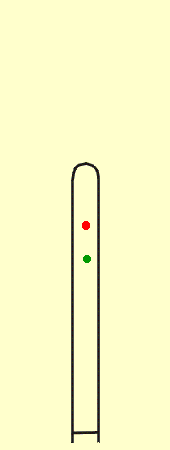
To understand development in the Basidiomycota you may wish to refer back to the diagram illustrating meiosis in the Dikarya. Unlike those of the Ascomycota, the four nuclei in the Basidiomycota do not remain inside the cell where they divided. Instead they migrate to the tips of horn-like protrusions called sterigmata (singular: sterigma). The tips of the sterigmata enlarge to form solitary basidiospores, each with a nucleus inside. The basidiospores then become separated from the sterigmata by their own walls and can be dispersed. Compare this with the animation of ascus-formation.
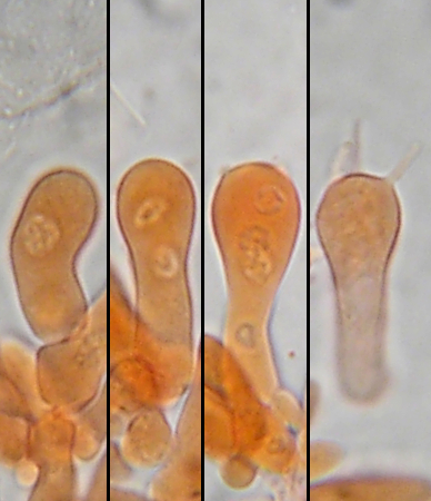
The pictures at right show four developing basidia, having 1-, 2- and 4 nuclei, plus one fully developed and empty basidium that had transferred its nuclei to the basidiospores, which were then discharged. The animation at far right shows how these stages progress. The photos showing 1- and 2-nuceate basidia could ones where nucear fusion and early meiosis had not yet taken place, but the size of the nuclei suggests that these basidia contain a fusion nuceus followed by the two nuclei from the first meiotic division.
Purely as an aside, it is quite unusual to observe nuclei in fungi with ordinary microscopic mounting media. The basidia shown here were found on the gills of very young fruiting bodies of the mushroom Gymnopus dryophilus mounted in KOH plus the stain Congo Red, a dye commonly used by mycologists.
Basidiospores are usually ejected forcibly from the basidium. However, unlike ascospores they do not travel very far, usually clearing the sterigata by only a few micrometers (thousandths of a millimeter). From there they are at the mercy of wind currents. Because of their small size and lightness they may travel great distances before settling. The mechanism of basidiospore discharge is less well understood than it is in the Ascomycota but is thought to involve minute changes in surface tension or electrostatic charges associated with a small drop on the sterigma just below the point of spore attachment. The relationship between spore and sterigma must be precise and it apparently allows for little variation, even between very distantly related species.
While the relationship between spore and sterigma remain fairly constant the overall form of the basidium can vary quite a lot. These variations in basidium structure are of great importance in classifying members of this subphylum and are discussed in more detail in the section on classification of the Basidiomycota.
The picture below illustrates a section of the hymenium lining the lamellae (gills) of Gymnopilus ventricosus a mushroom decaying the wood of conifers on the west coast of Canada and the United States. The hymenium is a palisade of basidia interspersed with some sterile cells.
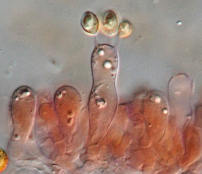 At the centre is a basidium with three sterigmata and young basidiospores visible. A fourth spore is present but out of focus. The red colour of the spores is caused by a reaction of their walls with Congo Red
In common with most mushrooms Gymnopilus ventricosus is able to eject its basidiospores forcibly from the sterigmata. Since the basidia make up the hymenium lining the surface of the lamellae, the discharged basidiospores find themselves suspended in the air between the lamellae. The air in this space is quite still, so the basidiospores fall out from the mushroom to be carried away be wind currents. This phenomenon is easy to demonstrate; simply place the cap of a mushroom on a sheet of paper, cover it with a bowl, and leave it overnight. By morning the paper will bear a mass of spores called a "spore print" mirroring the pattern of lamellae on the cap.
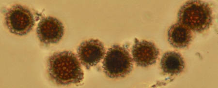
Some Basidiomycota do not forcibly eject their basidiospores from the sterigmata. In these species the spores are released when the basidium disintegrates. This is a common occurrence among Basidiomycota and is best demonstrated in the fruiting bodies called "puffballs". Puffballs are ball-shaped fungi with basidia packed tightly in their interior. The basidia finally disintegrate and the interior of the fruiting body is then filled with dry basidiospores. Raindrops or wind cause the delicate skin to break and then the spores puff out like smoke to be carried by the wind.
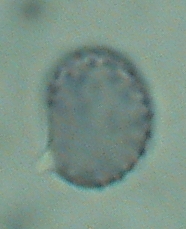 Basidia and basidiospores lacking forcible discharge can be recognized by their lack of certain features. First of all, the basidiospores are attached firmly to the sterigmata and are not easily dislodged before maturity. The attachment of the sterigma to the apiculus of the basidiospore is broad and square. Secondly, basidiospores that are not forcibly discharged tend to be radially symmetrical, as compared to the bilaterally symmetric basidiospores of forcibly discharged species. The bilateral symmetry of forcibly discharged spores appears to play some role in their release from the basidium. The basidiospore at right is from Russula densifolia. It is a typical forcibly discharged basidiospore; this side view shows it to be asymmetrical and to have a prominent and rather pointed apiculus. The picture at left shows basidiospores of Scleroderma macrorhizon, a type of puffball that grows in sand dunes. These spores appear round in any view because they are radially symmetrical. No matter how many
 spores are examined they will always appear circular. Non-forcibly discharged basidiospores are not necessarily spherical like those of S. macrorhizon. They may also be egg-shaped or even cylidrical, as in some stinkhorns, but they are always radially symmetrical.