Fleshy Fungi of New Brunswick >>
Pholiota spumosa
A Focus on Species >>
Pholiota spumosa
Pholiota spumosa (Fr.) Singer
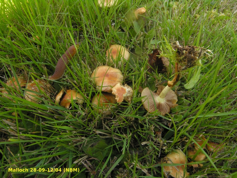
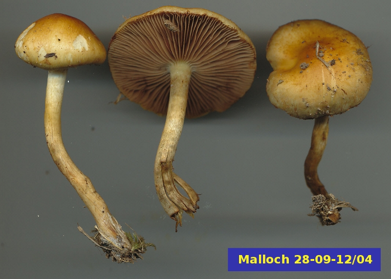
Pholiota spumosa is commonly found on Canadian lawns in the fall. Although it appears to arise from soil it actually grows on buried wood and roots. The picture on the title page shows its typical clustered growth habit among the grasses. In this picture, notice the slug that had been feasting on the caps of the mushroom, especially the thoroughly pruned one at centre. The pictures above show some fruiting bodies from the same location. The small one at right was still very young and retained the partial veil covering the gills. At maturity this veil material has mostly disappeared and may leave only a subtle trace of its presence on the upper part of the stipe.
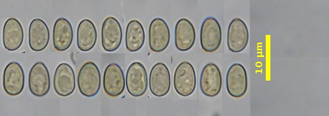
Members of the genus Pholiota belong to the family Strophariaceae. All species in this family have yellow-brown to purple brown basidiospres, seen clearly here as a "spore print" on the glass microscope slide. To the right of the slide is a group of these basidiospores viewed at high magnification. As you can see, they have quite smooth walls.
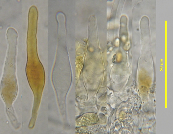
Another feature of the Strophariaceae found in most species of Pholiota is the presence of chrysocystidia, sterile microscopic structures occurring on the sides and edges of the gills. These cells are produced among the basidia on the gills. Their function is still not known, although many mycologists suspect they play some role in limiting the feeding of insects on the tissues of the mushroom. Chrysocystidia are characterized by their contents, which are golden yellow under the microscope. The ones in the picture here were stained in Melzer's solution and appear orange. As you can see, not all of them have taken up the orange stain. Strictly speaking, chrysocystidia should have a distinct body within them showing the yellow colour, but in fact many only show a more generalized reaction like the ones here.
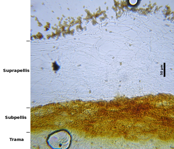
Pholiota spumosa, in common with many of species in this genus, may have caps that are slimy or viscid in wet weather. This slime is a gelatinous substance produced by the hyphae in the uppermost layers of the cap. The picture at right is of a cross section made through the flesh of a cap. It is made up of three layers, the suprapellis, the subpellis and the trama. The suprapellis is the part that becomes gelatinous and is entirely responsible for the slime. The subpellis is a solid layer below the suprapellis that serves as a sort of surface for the slime. The trama is merely the "flesh" of the cap and accounts for its thickness all the way through to the gills. The term "pellis", used with "supra-" and "sub-" in this example, simply refers to a skin-like tissue. When the pellis is on a cap mycologists call it the pileipellis; when it refers to the skin of the stipe it is called the stipitipellis
.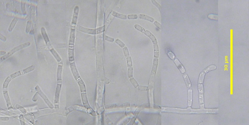
The picture at left doesn't look like a mushroom at all, but this is what you get if you try to grow P. spumosa in a Petri dish. The hyphae is this culture produced short branches, each of which became divided into individual spores or conidia. Unlike the production of basidiospores, conidia are the result of asexual reproduction. When they find a suitable place to grow they germinate to form a clone of the parent. In common with many other mushrooms, conidia of P. spumosa retract their contents toward the centre, leaving a small empty area between the next conidium. The empty area is separated by a septum so that the area between conidia often appears to have three walls. This type of cell separation is termed rhexolytic by mycologists. When we see it we are almost certain that were are looking at a basidiomycete. It is not always easy to see, but some hyphae, such as those at the left side of the picture, will oftem show it very clearly.
Photo: D. Malloch