A Focus on Species >> Dothiora pyrenophora
Dothiora pyrenophora (Fr.) Fr.
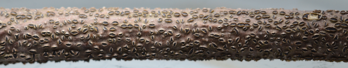
Dothiora pyrenophora (Dothideales: Dothioraceae) is one of many small ascomycetes found on recently dead branches and twigs. In common with many fungi occurring in this habitat it probably grows within the living tissues of its host and remains there harmlessly as an endophyte. The picture above is of a dead twig of mountain ash (Sorbus decora) collected in late March in southern New Brunswick. The twig, about a centimeter in diameter, was found on a small plant that had been damaged by a porcupine. Although dead, the twig was still partially attached to the living parts below.
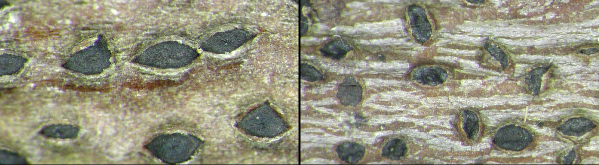
The two pictures above show ascomata of Dothiora pyrenophora in their natural habitat. The one on the left was taken in March when the weather was still quite wintery, while the one on the right was taken in May. Both were collected in the same general area as the twig shown in the picture at top. The ascomata in the March collection are smooth, and their contents were just reaching maturity. Those collected in May already show signs of senescence, with eroded and sunken tops. The hyphae of the fungus were inside the plant when it was alive and began to initiate its reproduction when the plant died. The early stages of ascomatal development would have occurred below the bark, but as the ascomata enlarged they broke their way to the surface.
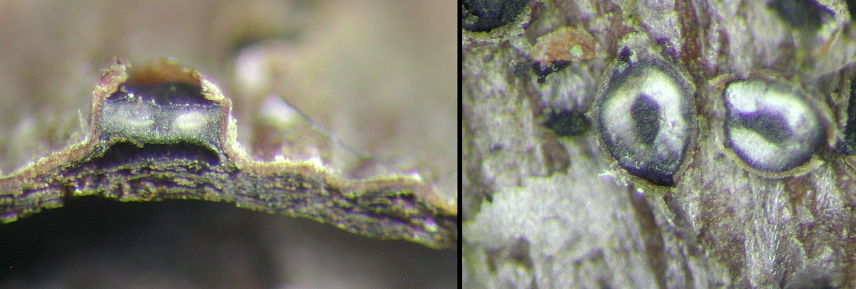
The next pair of photos shows two ascomata sliced open with a razor blade. At left, the ascoma has been cut through like a slice of bread, a longitudinal section. The result looks a little like a threatening face, with two eyes and a down-turned mouth. The two "eyes" are the parts of the ascoma bearing the asci, separated by a central sterile column. The photo at right is of a cross section of the ascoma, a sort of scalping, showing the contents to be in a circular configuration. The circle in the right part of the panel is the "lid" that was removed from the ascoma. If you think of these ascomata as containing a doughnut of asci you will see the two sides in cross section (doughnut cut in half vertically) or a full circle (doughnut cut in half so that each person shares the hole). Very few fungi have these characteristics.
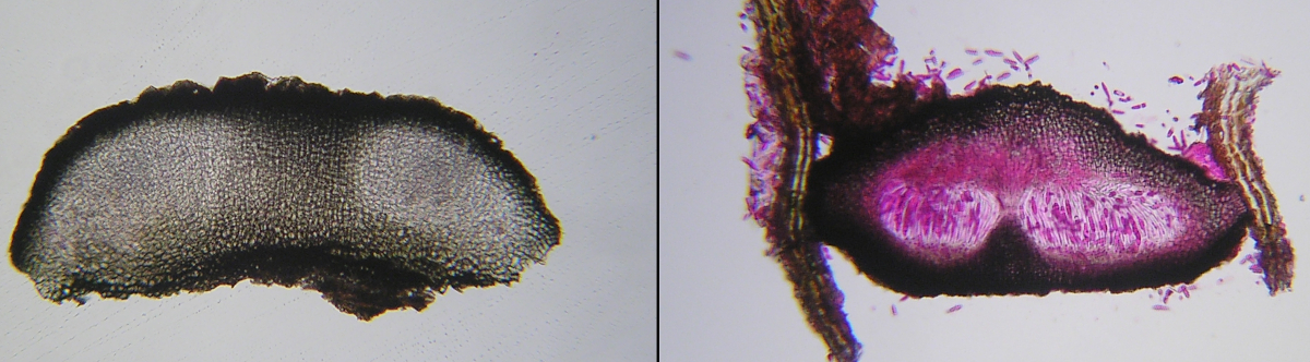
When they are still immature, the ascomata are quite solid, as seen in the microscopic view above left. The interior is composed of block-like cells forming a quite solid texture. However, even in that view we see the two light regions that will become the ring of asci and the dark central column that will remain mostly unchanged until the spores are released. The picture above right is of an ascoma that was sectioned two months later and dyed with aqueous Phloxine to better show the fertile regions. Here you can see the deeply-stained ascospores arranged more or less in rows in the vertically-oriented asci. The central column is quite narrow in the picture, not because it has disintegrated, but because the whole section of the ascoma was probably not cut exactly through its centre.
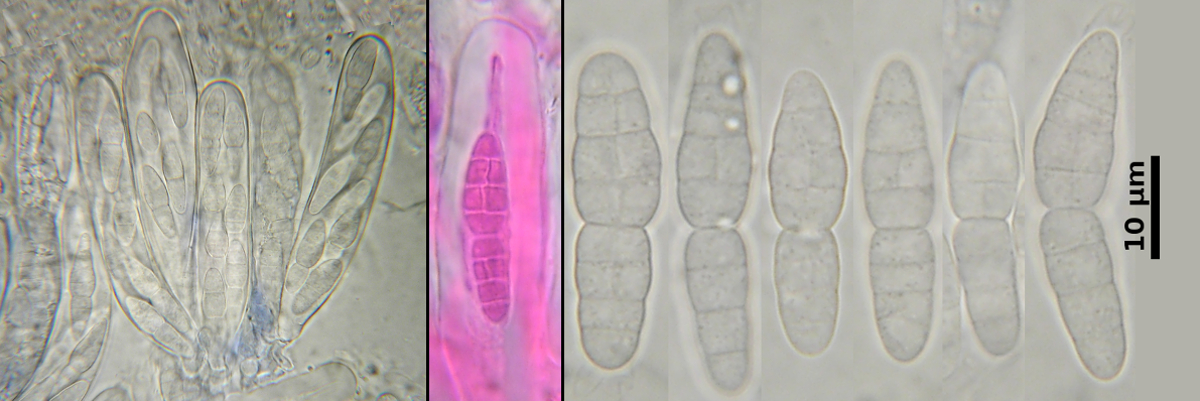
The asci, in common with those of other Dothideales are bitunicate. You can see this best in the middle panel above, stained with Phloxine, where the inner part of the ascus is stained pink while the outer layer remains more or less unstained. The ascospores, above right, have an upper cell that is broader than the lower one. They are muriform, meaning they have both transverse and longitudinal septa. They are strongly constricted at the middle or primary septum and may sometimes come apart here to form two distinct units.
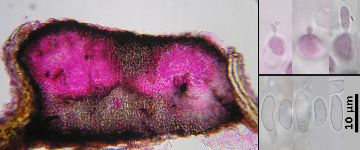
Dothiora pyrenophora also reproduces asexually in the form of large conidiomata resembling the ascomata. Unlike many ascomycetes it produces sexual and asexual structures at nearly the same time, so that it's actually necessary to cut one open and examine it microscopically to determine which kind it is. The photo above left is of one of these, also stained with Phloxine. Instead of containing asci and ascospores as deiscussed above it contains large numbers of conidia. Closer examiunation shows these to be produced on broad phialides, shown above right at top, produced around the wall of the conidioma. The conidia. above right at bottom, are irregular in shape but often resembling small beans or sausages.
Photo: D. Malloch