DISCUSSION OF THE DOTHIDEOMYCETES

The Dothideomycetes form a large and diverse group of species found in all parts of the world where fungi can grow. They are fairly well-defined and relatively easy to recognize. The core features of the Dothideomycetes, illustrated at left, are their ascostromatic development and their bitunicate asci.
Ascostromatic development
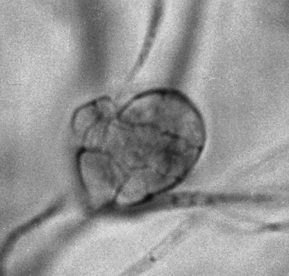
Ascostromatic development is a phenomenon that has been known for more than 100 years but its significance was not widely appreciated before the appearance of Dr. E. S. Luttrell's insightful monograph "Taxonomy of the Pyrenomycetes" in 1951. To grasp this concept, first go back and review our discussion of development in the Sordariomycetes. There it is explained that members of the Sordariomycetes have perithecia that begin as small coiled or otherwise characteristic hyphae, one cell of which functions as the ascogonium, the cell that receives a compatible nucleus during sexual reproduction. The Dothideomycetes differ from this model in having perithecia initiated by purely non-sexual vegetative means. This vegetative process begins as a simple division of a hyphal cell, usually in several different planes, to form a multicellular and more or less solid stromatic body, such as the one produced by Preussia cylindrica in the photo above left. This simple stroma will eventually begin to form asci inside following some kind of fertilization event. The stroma may be small and contain one to several asci or it may become much larger and contain many separate groups of asci. There is never a "true" perithecial wall initiated by the sexual process as in the Sordariomycetes. If you break one of these structures open it may resemble the perithecium of the Sordariomycetes, but it will never contain a distinct perithecium as happens in the Sordariomycetes. Some authors have restricted the use of the term "perithecium" to the ascogonium-initiated structures produced by the Sordariomycetes and have called the ones produced by the Dothideomycetes "ascostromata".
Bitunicate asci


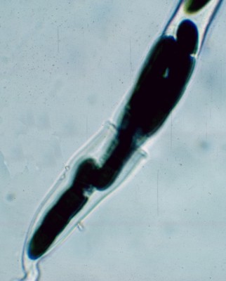
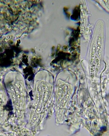
The other defining feature of the Dothideomycetes is the bitunicate ascus. Unlike asci in the Sordariomycetes and Leotiomycetes those of the Dothideomycetes have a thick double wall without any apical pores. These asci have a complex form of dehiscence whereby the rigid and brittle outer wall breaks, allowing the flexible inner one to extend out and forcibly discharge its ascospores. Because an inner structure bursts forth from an outer one this kind of ascus has been called the "jack-in-the-box" type. In the picture at far left an ascus of Preussia minima has had its outer wall broken, allowing the inner one to extend. The outer wall is the vase-like part toward the base of the ascus and the inner wall is everything above its rim. The very dark objects inside the ascus are the four-celled ascospores. The asci in the photo at near left are of a species of Arthonia. Three of the asci are still unexpanded and show the thick wall typical of the Arthoniales. The rightmost of these asci has broken to allow the inner wall to expand. There is some variation in how bitunicate asci open and eject their spores; our explanation here is admittedly a simplification.
Some terminology
The Dothideomycetes are a difficult group to come to terms with. There are many families differing in a variety of characteristics. Before proceeding further is will be useful to define some terms.
Ascoma (plural: ascomata), ascocarp
These are general terms denoting the ascus-bearing structures of the Ascomycota. Cleistothecia, perithecia, apothecia, hysterothecia, etc. are all types of ascomata. We follow current usage in preferring the term ascoma to ascocarp, but the latter is the one you will find in all the older (but still useful) literature.
Centrum, carpocentrum
The inside of an ascoma where the asci are produced.
Hamathecium
Tissues surrounding the asci within the centrum of the ascoma. These can take several forms, highly important in recognizing the orders and families of Dothideomycetes
Interascal pseudoparenchyma - the original and unchanged tissue of the centrum. In this type the asci are simply separated from one another by simple unmodified tissues.
Paraphyses - hyphae arising from the base of the centrum and growing toward the centre or apex. Typical of discomycetes and Sordariomycetes.
Pseudoparaphyses - hyphae arising at the top of the centrum and growing down to become anchored at the base. They may later become detached at the top of the centrum and come to resemble paraphyses. It may be necessary to examine razor-blade sections of young ascomata to be certain of this distinction. Two types of pseudoparaphyses have been recognized:
Cellular pseudoparaphyses - pseudoparaphyses that are broad and clearly septate. The septa can be seen at 400X under the microscope. They may be numerous enough to support one another or be more separate and embedded in a gel.
Trabecular pseudoparaphyses - pseudoparaphyses that are thin, branched and only sparsely septate. The septa cannot been seen at 400X magnification. They may be crowded or distant and are nearly always embedded in a refractive gel.
Paraphysoids - tissues occurring between the asci that may become stretched and resembling pseudoparaphyses. Unlike pseudoparaphyses they do not arise from the top of the centrum and grow down; they are formed in-place. Paraphysoids are particularly characteristic of Dothideomycetes forming apothecial ascomata.
Periphysoids - Similar to pseudoparaphyses in arising at the top of the centrum and growing downward. They may be very short and never become anchored at the base of the asci.
No hamathecium - some asci are borne in the centrum without any surrounding tissues.
The Dothideomycetes are still only poorly understood. The class has been divided into several orders but these are not uniform from author to author and are not confirmed by genetic studies. The feature that has been most used in the modern literature to subdivide the Dothideomycetes is the hamathecium. This may appear at first to be the most troublesome and arcane means of accomplishing this, but it does work. Usually the perithecia or ascostromata occur in large umbers and it is not difficult to find both immature and mature examples. You then must take a sharp razor blade and cut a thin section of each, rather like a slice of bread from a loaf, and mount it on a microscope slide for examination. Of course this requires the intermediate magnification of a binocular dissecting microscope to accomplish, so if this is not available you will just have to make do as well as you can without. Sometimes a simple squash mount will be enough to confirm the presence of a hamathecium.
Using the hamathecium and other features as a guide we can recognize the following orders:
MYRIANGIALES - Centrum with interascal pseudoparenchyma. The asci are well separated from one another or appearing to be solitary. The ascomata are often irregular in shape and not forming recognizable perithecia (includes the Seuratiaceae)
ARTHONIALES - Centrum with tightly packed paraphysoids. The balloon-like asci form a continuous hymenial layer. The ascomata are often irregular in shape, flattened and open by the irregular break-up of the covering layer
PATELLARIALES - Centrum with paraphysoids. The ascomata are apothecial at maturity
PLEOSPORALES - Centrum usually with pseudoparaphyses that may or may not be trabiculate
JAHNULALES - Centrum with pseudoparaphyses. Ascomata are typical perithecia. There are only three genera in the Jahnulales, Aliquandostipite, Jahnula and Patescopora. All species occur on decomposing plant materials in fresh water. They are similar to members of the Pleosporales, but molecular studies have shown them to be distinct.
HYSTERIALES - Centrum with pseudoparaphyses. The ascomata are hysterothecia, elongated structures that open by a slit rather than an ostiole
CAPNODIALES - Centrum mostly with no hamathecium although some members of the family Metacapnodiaceae have periphysoids extending a short way down from the top. These grow as "sooty moulds", black mycelial coverings of trees and shrubs
DOTHIDEALES - Centrum with no hamathecium. Asci occur in clusters with no accompanying tissues. Ascospores with more than one cell.
BOTRYOSPHAERIALES - Centrum with or without hamathecium. Asci occur in clusters amongst pseudoparaphyses, but these often dissolve before the asci mature. Ascospores one-celled.
It should be understod that there are undoubtedly more than seven orders in the Dothideomycetes. Myconet, the online publication listing all known ascomycetes, lists 24 families within these seven orders but lists another 44 families that have yet to be assigned to any order. From this we can conclude that 65% of the Dothideomycetes are still so poorly studied that we cannot classify them with any confidence. There is still much to be done.