LAMELLAE, PORES OR TEETH
FORM
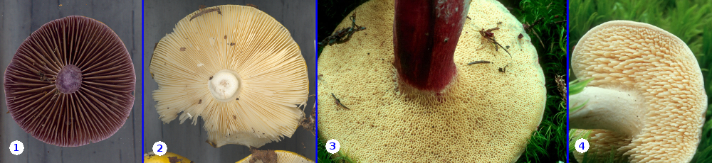
The main function of a mushroom is to disperse its spores into its environment, thus ensuring the establishment of offspring. The spores are produced by basidia which are themselves anchored to a variety of surfaces or structures called the hymenophore. The most common types of hymenophore in mushrooms are lamellae (gills), pores and teeth, ass illustrated in the above set of photos. The lamellae of Cortinarius evernius (1) are the most typical form. These radiate from the stipe out to the margin of the cap like spokes on a wheel. As they radiate the become more distant from one another and add new ones to fill the gap. These small lamellae that do not originate at the stipe are called lamellulae, meaning "little lamellae". In many species of Russula, such as R. claroflava (2) the lamellae radiate from stipe to edge of cap without adding lamellulae. Any large mushroom lacking lamellulae is almost certain to be a species of Russula.
The two panels to the right are of Xerocomus subtomentosus, a bolete and Hydnum repandum, a typical tooth fungus. The hymenophore in boletes is produced as long tubes lined with basidia while in tooth fungi the basidia are borne on the outside of elongated teeth.
ATTACHMENT
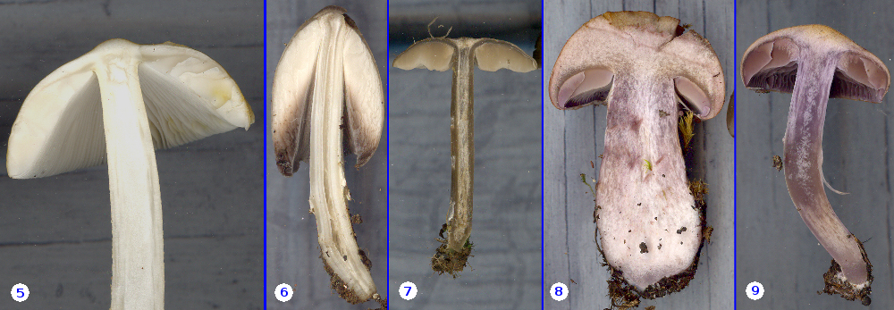
One of the fundamental features of mushrooms that can be used to identify them is the way in which the lamellae are attached to the stipe. These can be:
Free - The stipe is structurally distinct from the pileus, often with a ball-and-socket relationship. The lamellae are not attached to the stipe, but instead arise from the tissues of the pileus. Free lamellae are less common than attached ones but are characteristic of several genera such as Agaricus, Amanita, Coprinus, Coprinopsis, Macrolepiota and several others. In the set above, Panel 5 is a photo of Amanita sp. and Panel 6 is Coprinopsis atramentaria.
Adnexed - Here the lamellae are ascending at the point at which they meet the stipe. In Panels 7 and 8 the lamellae of Entoloma sericeum and Cortinarius sp. are so deeply ascending that they almost appear to be free. However, the stipe and the cap are of very similar and intergrading tissues so that we cannot really say the lamellae are not entirely free of the stipe as they would be in the ball-and-socket type. Panel 9 is of Cortinarius evernius. Here the lamellae are just barely ascending and might be classified as our next type, adnate. In making a description of a mushroom this is not a big problem, we just describe the lamellae as "adnexed to adnate".
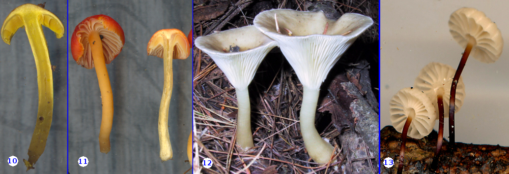
Adnate - the lamellae appear to meet the stipe a nearly a right angle and are neither ascending nor descending. The photo in Panel 10 is of Cortinarius croceus.
Decurrent - Decurrent lamellae appear to run down the stipe. This is diagnostic for such genera as Clitocybe and Cantharellus. Hygrocybe mucronella, in Panel 11, has lamellae that are clearly ascending but which end up being decurrent. Ampuloclitocybe clavipes in Panel 12 shows a more straight-forward decurrent condition.
A collarium - the tiny Marasmius rotula is the best example of lamellae with a collarium. Here the lamellae never meet the stipe, but instead form a little collar around it.
SPACING
The spacing between the gills of a mushroom varies quite a bit from species to species and may be useful in identifying an unknown. Describing gill spacing in a useful way is more difficult. Because the gills radiate out from the stipe they start out very close together and are much further apart at the edge of the cap. Along the way they may or may not produce lamellulae to fill the increasing space. Some mycologists have tried to measure the distance between gills at the margin of the cap or to count the number within a pie-shaped piece of cap of known angle. Most people find these methods cumbersome and soon abandon them.
A more convenient, although imprecise, method is to place gill spacing into a few catagories. The most commonly used of such catagories are "crowded", "close" and "distant". These are often refined further by using the prefix "-sub" with each of the three terms. The problem with "-sub", though, is knowing exactly what the term means. Are gills that are called "subclose" closer then "close" or more distant than "close"? Given the lack of agreement on this subject it is probably best just to be consistent in your use of the terms. Of course a photograph of the gills settles the issue at identification time. Here is the system I use in my notes:
Crowded
The gills are so close together you may have trouble seeing the spaces between them. This is typical of young speciemns of the market mushroom Agaricus bisporus, many species of Amanita (Panel 5) and Coprinopsis (Panel 6).
Close
Close gills are fairly densely arranged, but the spaces between them are easily seen. Russula claroflava (Panel 2) and Ampulloclitocybe clavipes (Panel 12) are a good examples.
Subclose
This is probably the most common situation in mushrooms. The gills of Cortinarius evernius (Panel 1) are representative.
Distant
Many species of Marasmius are tiny mushrooms with gills that are very widely separated. Marasmius rotula in Panel 13 is quite typical. Bear in mind, that these mushrooms are really small and that their gills, in millimeters, are no further apart than those of large mushrooms stated to have close gills. It's all relative to size.
Many of the mushrooms you collect, such as Hygrocybe mucronella (Panel 11) will have gill spacing that seems to fall somewhere between these four catagories. No problem; just say so in your notes by writing "subclose to distant" (for Hygrocybe mucronella), "close to crowded", etc.
Boletes and tooth fungi present other challenges. For boletes and other fungi with pores it is conventional to express pore size as the number of pores per millimeter. For tooth fungi the length and diameter of the teeth may be of some importance.
MARGIN
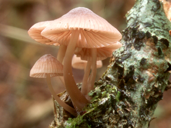
Although most mushrooms will have an even margin or edge, some will show interesting variations. In many genera the edges of the lamellae produce very few basidia and are instead lined with cheilocystidia, cells with distinctive shapes and colours. The microscopic anatomy of mushrooms is not the subject of this essay, but if you are curious, our illustrations of Tubaria confragosa show some photos of cheilocystidia in that species. If the cheilocystidia are abundant and filled with a coloured pigment the edge of the gill itself will have a different colour than the sides. Mycena rubrimarginata, pictured at right, is a small mushroom with gills that are nearly white, but red at the margin. Some species, such as those of Panaeolus species, may have coloured gills with white margins
Aside from differences in colour, the margins of the lamellae may appear ragged or fringed if the cheilocystidia are elongated or may appear to be serrated like miniature saw blades, a feature typical of Lentinellus species.
![]()