Fleshy Fungi of New Brunswick >>
Tubaria confragosa
Tubaria confragosa (Fr.) Harmaja


Two collections:
-
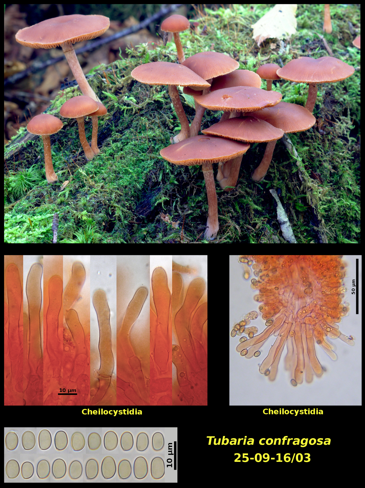
1. Clustered (many) on a decayed conifer log, Campobello Island, New Brunswick (25-09-16/03).
-
Pileus convex-hemispherical at first, expanding to broadly convex, without an umbo, striate at the margin, dark red brown (HSV20:70:60-70), hygrophanous, glabrous, moist, 12-38 mm in diameter. Stipe clavate at first, later equal although retaining the basal swelling, pale brown (HSV40:20-30:90), dry, glabrous or finely fibrillose, with an evanescent annular zone, 20-38 X 2-4 mm. Lamellae light orange brown (HSV30:30-40:90), subclose, adnexed, not marginate. Flesh conclourous with the surface tissues in the pileus and stipe, lacking a distinctive odour.
Basidiospores 5.8-7.7 X 3.9-5.1 μm, Q = 1.32-1.61 (average[20]: 6.6 x 4.5 μm, Q = 1.45), smooth. Cheilocystidia 37-66 X 6.5-8.9 μm, more or less cylindrical but somewhat irregular in diameter, abundant and forming a continuous sterile palisade. Pleurocystidia lacking.
-
-
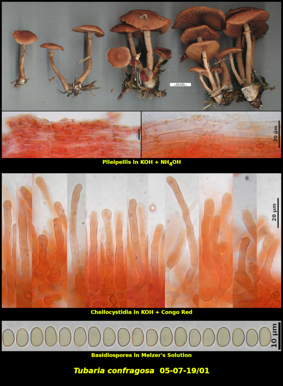
Pileus not seen when young, convex, slightly depressed at the centre, without an umbo, very obscurely striate at the margin, dry, glabrous, light red brown (HSV15:40-50:70), hygrophanous, 33 mm in diameter. Stipe equal, with a silvery white cansence overlying a pale reddish brown (HSV10:15-20:90) ground colour, dry, pruinose at the immediate apex and glabrous below, annulate, 42 X 3 mm. Lamellae red-brown (HSV20:40:80), adnate, close, not marginate. Annulus silvery white below, ascending, membranous, persistant. Flesh olivaceous (HSV35:15:70) in the pileus, concolorous with the surface tissues in the stipe, lacking a distinctive odour and taste.
Basidiospores light orange brown in spore print, ellipsoidal to slightly ovoid, sometimes slightly phaseoliform in profile, smooth, darkening slightly in Melzer’s Solution but neither amyloid nor dextrinoid, 5.8-7.4 x 4.2-4.7 μm, Q = 1.36-1.63 (average[33]: 6.8 x 4.5 μm, Q = 1.50). Cheilocystidia forming a continuous sterile margin, filiform to sublageniform, 32-87 x 4.9-7.5 μm. Pleurocystidia lacking. Pileipellis a simple cutis of repent, radially encrusted hyphae. Basidia 4-spored, clamped at the base.
2. Clustered (many) on a fallen and decaying log of Betula papyrifera in forest dominated by Abies balsamea, Betula papyrifera and Picea mariana. Upper Dungarvon Protected Natural Area, New Brunswick (05-07-19/01).
In common with other species of Tubaria, the basidiospores of T. confragosa are quite pale under the microscopic and often collapsed, especially in water mounts. The ones in the photograph were mounted in Melzer's Solution and heated gently to make them pop back into their normal shape.
Although the two collections presented here appear to be nearly identical, both macro- and microscopically, they differ in two features that are considered to be important in the taxonomy of other genera: i) date of appearance and ii) substrate. Collection 25-09-16/03 was collected on the wood of a coniferous tree in late September, while Collection 05-07-19/01 was growing on a birch log in early July. However, two other collections of this species were also made in early July, in an area close to the site of Collection 05-07-19/01, one on coniferous wood and the other on birch. It may be that T. confragosa is just not that particular about what kind of wood it will decay.
Photograph: D. Malloch (25-09-16/03, 05-07-19/01).