Home >> What fungi are >> How fungi reproduce >> Sexual reproduction >> Sporangia
FUNGI REPRODUCING SEXUALLY BY MEANS OF SPORANGIA AND ZYGOSPORANGIA
SPORANGIA
Among the true fungi only the Chytridiomycota and Zygomycota reproduce sexually by means of sporangia. The Chytridiomycota are themselves a diverse group of fungi. They may have simple life histories with zygotic meiosis or, as in the mosquito parasite Coelomomyces, complex diplobiontic cycles. In all cases reproduction is by means of sporangiospores, spores borne inside sporangia that were the site of meiosis of diploid nuclei.

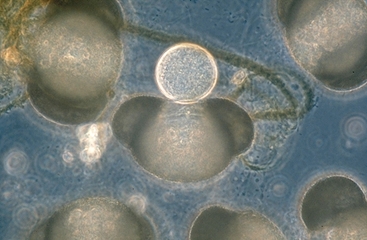
The photo at left shows a single sporangium of Chytridium sp. developing on a pollen grain of red pine floating in pond water. Pine pollen, along with that of fir, spruce and many other conifers, is characterized by having two large air bladders on one side, giving it the appearance of having ears. A spore of Chytridium sp. has swum in and settled between the "ears", where it has produced a sporangium. The sporangium would have gone on to produce internal spores. When released the spores can swim actively in the water by means of whip-like flagella. Simple chytrid sporangia like this one can produce sexual or asexual spores, depending upon whether the spore that produced them was haploid or diploid. More complex chytrids may have multiple sporangia connected by hyphae, but their appearance is usually much like this one.
ZYGOSPORANGIA
Sexual reproduction in the Zygomycota involves the production of zygosporangia and zygospores. This type of reproduction is fairly simple and is related to conjugation in certain algae. Mating is initiated when two hyphae called gametangia become connected by a short bridge. The central part of the bridge receives nuclei from both gametangia after which it is separated from both by septa. Compatible nuclei within this new cell fuse to form zygotes and then immediately or after some delay undergo meiosis to bring about a reassortment of the parental chromosomes. The cell where these events take place becomes surrounded by a thick wall and becomes a resistant spore-like structure called a zygosporangium. Usually the outermost layers of the zygosporangium wear away or disappear and the remaining body is called a zygospore. At an appropriate moment the zygospore may germinate to produce a new thallus or it may give rise to asexual spores via a sporangium.
 |
 |
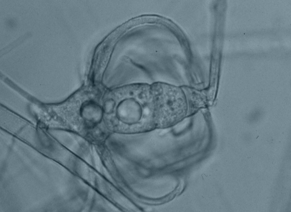
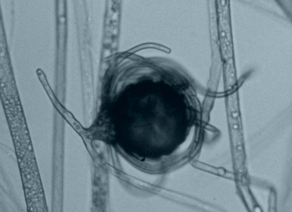
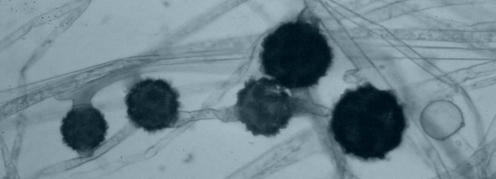
The two pictures above show zygospores of Absidia spinosa, a common fungus in soil. The one at left is at an early stage of development where the zygosporangium has been partially deliminated by septa but still retains its light colour. The picture at right shows an older zygosporangium that has a rough dark spore wall. The curved branches surrounding the zygosporangia are called suspensor appendages. These appendages are diagnostic features of Absidia species. The picture below illustrates several mature zygospores of Zygorhynchus molleri, another common soil fungus. These zygospores are similar to those of Absidia spinosa but lack the suspensor appendages
