Home >> Diversity and classification >> True fungi >> Zygomycota
ZYGOMYCOTA
The Zygomycota take their name from their method of sexual reproduction, involving the production of conjugating gametangia, leading to the production of zygosporangia and zygospores. This is a relatively simple kind of reproduction found in a variety of filamentous organisms. Asexual reproduction in this group can be far more complex, at least structurally so, making it the primary means for identifying members of this group.
Before plunging into the details of these fungi it should be pointed out that their claasification is incredibly complicated. As more and more genetic evidence becomes available is is clear that what we call the Zygomycota is really a collection of several phyla, some more closely related to one another than others. In fact, some "Zygomycota" are more closely related to the Dikarya than they are to other "Zygomycota"! A detailed dicussion of this is well beyond the scope of these pages where the focus is mainly on life histories.
The fundamental asexual structure in the Zygomycota is also a sporangium, but the variety of these structures is quite amazing. Zygospores are usually thick-walled resistant structures capable of preserving the fungus over long periods of dormancy. Asexual sporangia, on the other hand, play a key role in the day to day lives of these organisms, allowing them to spread and colonize new substrates quickly and efficiently.
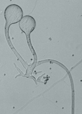
The affectionate couple at left are two sporangia of Rhizopus domesticus arising from a small nest of rhizoids. The root-like rhizoids help the sporangia to remain anchored to whatever object they may encounter. The thread extending to the right and down is a stolon, a hypha that grows out until it encounters another appropriate place to attach rhizoids and build sporangia.
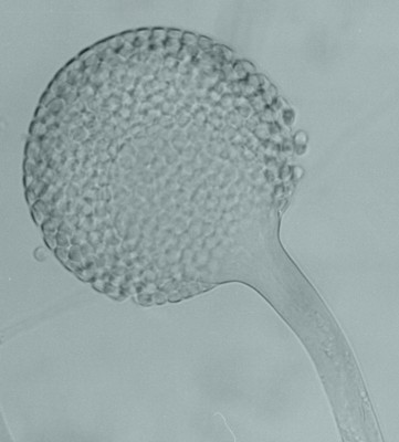 Stolons, rhizoids and sporangia all characterize the genus Rhizopus. The sporangia should contain spores, but they are either still immature or, more likely, have already dispersed their spores and remain now only as sporangiophores (the stalks) and columellae (the swollen apical parts). The smaller picture immediately to the left is of a single sporangium, also of Rhizopus domesticus. It is easy to see here that the spherical sporangium contains a large number of spores. But look more closely: inside you will see the very faint outline of the balloon-like columella extending into the spore mass. When the sporangial wall breaks, as it had done with the couple at far left, nothing is left but the columella and a few spores that were left behind. This is the normal type of sporangium as seen in Rhizopus, Mucor, Absidia, Circinella and other members of the family Mucoraceae. If you attempt to grow fungi in petri dishes you will soon encounter members of the Mucoraceae. They are found everywhere and are invasive and aggressive.
Zygomycetes are interesting fungi that are able to hold their own in the face of competition from members of the much larger Dikarya. They are characterized by several features that set them apart. First of all their hyphae are coenocytic, meaning that they have no septa. With no cross-walls to slow the flow of cytoplasm these organisms can extend their hyphae rapidly into new territory. They do not respond as well as the Dikarya to cellulose, sucrose and other common plant products and instead often seem to associate with materials of animal or fungal origin. They can be strong digesters of protein and even chitin, the fundamental material of insect exoskeletons and fungal hyphae.
Although the phylum is not large it is quite diverse. Several orders have been recognized. Many, such as the Mucorales discussed above appear to be saprotrophic and to respond most vigourously to simple sugars, such as glucose, and to amino acids and proteins. Other orders are characterized mainly by parasitic forms attacking fungi and animals. None appear to be parasites of plants, although Endogone pisiformis seems to be restricted to living mosses.
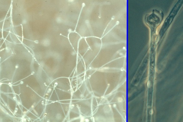
Rhizopus species and many Zygomycota produce sporangia with brittle walls and dry spores. When they are mature any air movement may be sufficent to carry the spores away to a new location. This is why Rhizopus species are a great pest in the laboratory. On the other hand many other Zygomycota, such as Absidia caerulea at right, maintain their sporangiospores in droplets of liquid. It is likely that these species do not use air currents for dispersal but instead depend upon insects and other small invertebrate animals to do the job. Absidia species are common in soil where minute animals are common but air currents are not. If you look carefully at the picture you will see the sporangia elevated above the hyphae and that each sporangium bears a drop of liquid. In the far right is a magnified view of a sporangium. here it can be seen that the sporangial wall has broken away to leave a cup-like structure at its base. This and the prominant columella inside help to maintain the spore-filled water droplet, unfortunately dissipated when the microscopic mount was made.
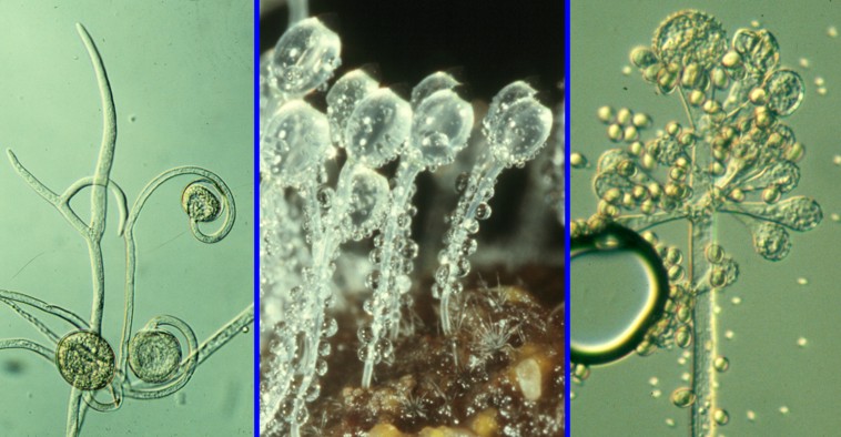
Not all Zygomycota have sporangia resembling those we have discussed so far. The picture above presents some variants. In the cell at left are sporangia of Circinella muscae, a fungus we sometimes encounter in soil or air samples. Here the sporangia are several on the same axis and each borne upon a coiled sporangiophore. The generic name Circinella refers to the fact that the sporangiophores are coiled or circinate.
The fungus in the middle is Pilobolus umbonatus a peculiar but very common fungus inhabiting piles of dung. The colourless sporangiophores are greatly swollen at the apex and are filled with water and cytoplasm under great pressure. The sporangia themselves are inconspicuous and can be seen as little black caps at the top of the swelling. The swelling functions in two ways, first it acts as a lens and focuses light down to light sensitive areas in the sporangiophore, causing it to bend toward the light. At maturity a thin spot at the top of the swelling under the sporangium breaks allowing a thin stream of water under great pressure to squirt out and propel the sporangium away. This is a powerful mechanism that can shoot the sporangium a meter or more. Quite a feat for a nearly microscopic organism. The top of the sporangium is sticky so that when it final completes its flight it will come down and stick to a blade of grass. A grazing animal may then come along, eat the grass and unknowingly pass the sporangium and spores through its digestive tract and back out on a newly formed pile of dung. Thus Pilobolus has a remarkable set of adaptations ensuring its dispersal from one dung pile to the next.
The rightmost photo is of sporangia of Cunninghamella elegans, a fungus usually found in soil in warmer climates. It differs from other species we have discussed in bearing numerous short branches terminating in a swelling and with each swelling producing external spores on its surface. Some mycologists have interpreted the swellings as columellae and each of the "spores" as an individual sporangium. In fact the spores do have a double wall, suggesting that the outer one is a sporangial wall and the inner the spore wall. Because of this complexity the "spores" are usually called sporangioles. The sporangioles are very lightly attached and are easily knocked off, as happened in the specimen in the photo. Here we have the swollen vesicles or collumellae, covered in tiny "teeth" where the spores were attached and many spores floating around beside them. Incidentally, the large black ring about halfway down the picture is just an air bubble on the microscope slide. You should ignore it.
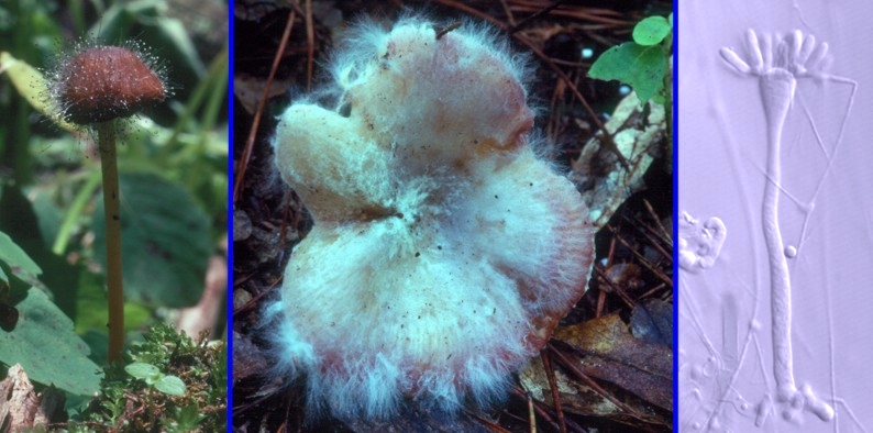
The set of photos at right represent another aspect of the Zygomycota, the mycoparasitic members. Many Zygomycota are active parasites of other fungi. Hosts can include other Zygomycota and members of the Dikarya. The leftmost panel is of Spinellus chalybaeus parasitizing a species of Mycena. The fungus has spread throughout the mushroom and is producing sporangiophores and sporangia out of the cap. In the middle panel a species of Russula has been attacked by Syzygites megalocarpus, one of the Zygomycota that essentially destroys its host. At later stages of infection the mushroom cap may be covered with zygospores. The fungus in the right panel is a species Syncephalis a group of mycoparasites infecting other Zygomycota. Species of Syncephalis are obligate parasites and cannot live in the absence of their hosts. The specimen in the photograph was found growing on a species of Mucor, itself growing on some decaying seaweed. Syncephalis also presents us with another variant on the zygomycotan sporangium. Here, as in Cunninghamella, the sporangiophore is enlarged at apex into a columella-like structure. Arising from this are several "fingers", each of which will become a miniature sporangium (a merosporangium) containing a few spores.
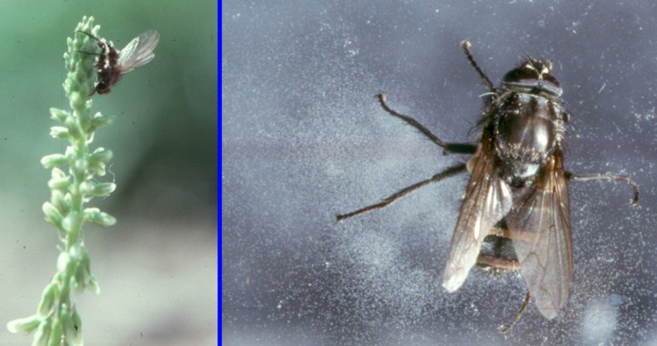
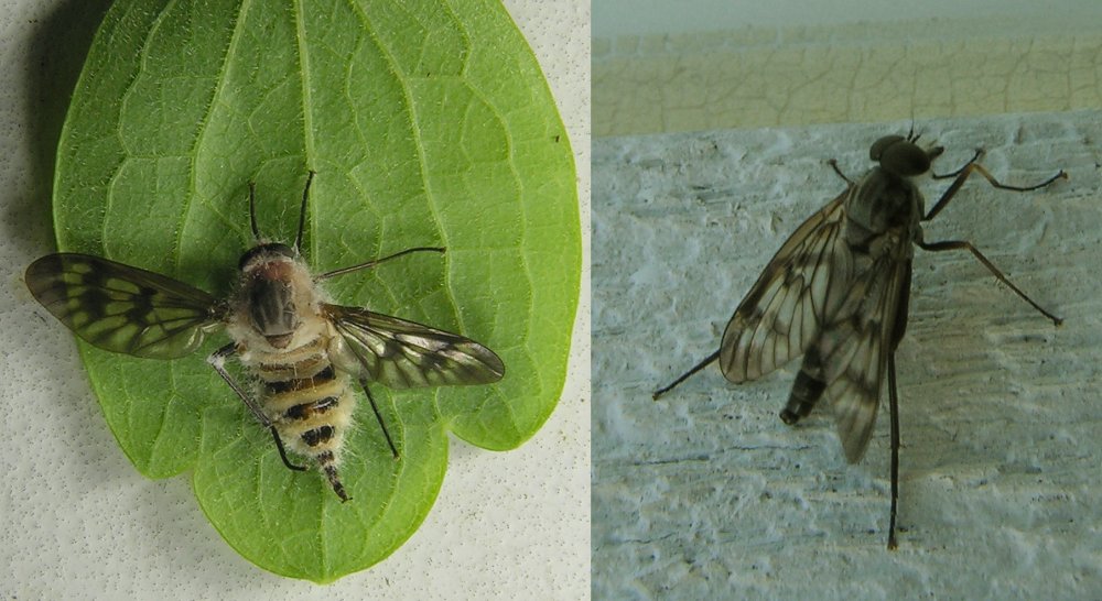
The Zygomycota can also act as parasites of insects. Above are two examples of this group infecting flies. Members of this group infect insects and completely fill their bodies with hyphae. Before the insect dies the fungus may cause it to perform one last act, positioning itself so the fungus will be in the best place for its dispersal. In the leftmost panel a fly infected with a species of Entomophthora has landed on a flower stalk, turned itself head-down and died. The fungus will then produce its sporangia out through the abdominal segments and forcibly shoot the spores into the air. It is still not known how these spores manage to strike another fly. One suggestion is that other flies will mistakenly attempt to mate with the dead fly and get covered with spores, but this is purely conjectural. The fly in the panel second to left, photographed by Dr. R. Greg Thorn at the University of Western Ontario, has died on a window pane. Here the white sporangial masses at each abdominal segment are clearly visible, as are the many spores adhering to the glass. The third panel shows a snipe fly, Rhagio mystacea, that has crawled beneath a leaf and has died. Its abdomen is swollen and is producing a large numbers of sporangia of Furia ithacensis. Dr. Kathie Hodge, in her Cornell Mushroom Blog, has an interesting discussion of this fungus. Mycologists attending the Bioblitz held by the New Brunswick Museum at Caledonia Gorge Protected Natural Area in June 2011 discovered many such infected snipe flies. The individual in the rightmost panel is healthy. Recent genetic studies have shown that Entomophthora and its relatives are rather distantly related to other Zygomycota. Traditionally they have been placed in their own order, the Entomophthorales, but they are now often elevated to the level of class (Entomophthoromycetes) or even phylum (Entomophthoromycota).
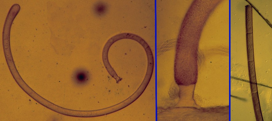
Insects and other invertebrate animals can also host members of the Zygomycota inside their digestive tracts. The species of Enterobryus pictured at left was found attached to the wall of the midgut of an American giant millipede (Narceus americanus) in central Ontario. Enterobryus is a simple member of the Xygomycota, consisting of a single filament extending out from the inner gut wall of the host and absorbing nutrients as they pass by. It is sort of a fungal equivalent of the tapeworm. The left panel shows the whole fungus, while the middle panel show the suction-cup-like holdfast that anchors it to the gut wall. The third panel shows the end of a filament that has been converted into a sporangium containing a row of spores. Enterobryus and its relatives belong to the order Eccrinales. Relevant to our opening caution about classification of the Zygomycota. studies by Cafaro (Molecular Phylogenetics and Evolution 35:21-24. 2005) have revealed that the Eccrinales are not fungi at all but members of the class Ichthyosporea. In spite of their non-fungal heritage they are still studied mainly by mycologists.
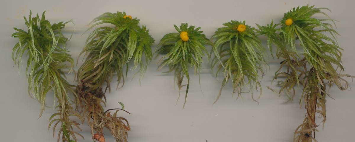
Finally, in the picture above, we come to a member of the Xygomycota that actually grows on living plants. The yellow objects at the top of the plants are produced by Endogone pisiformis, a species that apparently parasitizes mosses and produces bright yellow masses of thick-walled spores at the apex of the stems. The fungus is usually found on peat mosses (Sphagnum species) as in the picture here, but it can grow on other mosses as well.