Home >> Diversity and classification >> Fungus-like organisms >> Slime moulds
SLIME MOULDS
The term "slime mould" is used to described an array of organisms that have amoeboid stages in their life histories and with these amoeboid stages usually aggregating to form a plasmodium (plural: plasmodia) or pseudoplasmodium. Although these technical terms may seem a little intimidating at first they are a concise way of describing a very particular and interesting mode of existance. The term "amoeboid" refers to a cell that resembles an amoeba (plural: amoebae) , that is, one that has no cell wall and moves by "flowing" over its environment. Many amoebae extend their cells unequally in the form of arms or pseudopodia and are able to surround and enclose their prey.
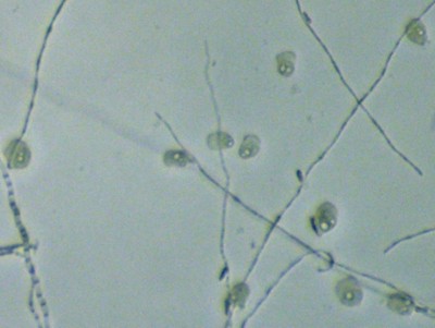
In the picture to the left ten amoebae are slowly picking their way among some fungal hyphae. Like most amoebae these are probably engulfing and digesting small organisms, particularly bacteria. The white structures visible within the amoebae are vacuoles, small sacks within the cytoplasm. The vacuoles are surrounded by a membrane through which certain substances can pass back and forth. Vacuoles may the the site of food storage, digestion and water relations. The particular amoebae in this picture were found on rotting seaweed on a Bay of Fundy beach. It is difficult to determine whether these amoebae belong with the group we call slime moulds because they are solitary, at least at this stage.
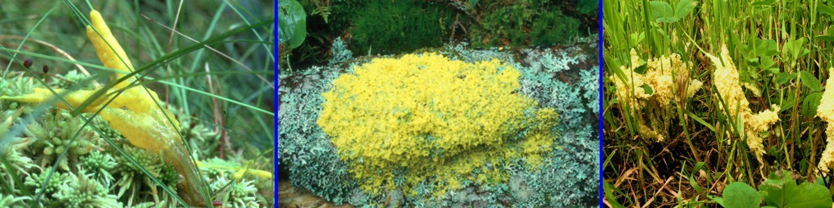
To be considered a slime mould the amoebae must first aggregate and then cooperate to form complex reproductive structures. Aggregation of amoebae can result in a true plasmodium, where individual cells come together and fuse to form one large multinucleate individual. Plasmodia, such as those of Fuligo septica illustrated in the three pictures above, can be up to several centimeters across and cover conspicuous expanses of soil or vegetation, even urban lawns where they may appear overnight to the consternation of fastidious homeowners.
In many cases the amoebae aggregate but maintain their individuality so that they do not form a true plasmodium. Such aggregations of individual amoebae are called pseudoplasmodia.
Whether forming a plasmodium or a pseudoplasmodium there would not be much reason to call these organisms fungus-like were it not for their reproductive stages. In pseudoplasmodial slime moulds the amoebae take on different roles within the pseudoplasmodium. Some begin to pile up upon one another and form cell walls, eventually producing a multicellular stalk. Other amoebae move up the stalk and become converted into a mass of spores or cysts at its apex. At this stage the organism looks like a fungus, superficially resembling the sporangia and sporangiophores of Mortierella or Mucor species (Zygomycota) or, if branched, one of the hyphomycetes like Verticillium. The stalk with one or more wet masses of cysts at its apex is called a sorocarp. These pseudoplasmodium-forming amoebae, called cellular slime moulds, are divided further into the Acrasiomycota and the Dictyosteliomycota.
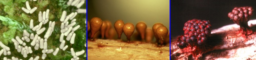
More familiar to naturalists are the true plasmodial slime moulds, often called myxomycetes and classified in the phylum Myxomycota. These organisms form plasmodia that spread over all manner of objects in their pursuit of bacteria, yeast and other small bits of organic material. Unlike the cellular slime moulds the myxomycetes form complex and often large spore-bearing structures containing enormous numbers of spores. These structures are generally recognized to take the form of sporangia, plasmodiocarps or aethalia. The three pictures above and three more immediately below illustrate some common sporangial myxomycetes. The sporangia are characterized by their individuality or separateness from adjacent sporangia. They may be stalked or sessile (unstalked). The species above are, left to right, Arcyria cinerea, Hemitrichia clavata and Metatrichia vesparium. Note that the sporangia of H. vesparium have become united together to form a compound structure but are clearly distinguishable as individual structures.
The next three species (above) are Stemonitis fusca, Diderma globosum and Trichia varia. Stemonitis fusca, one of our largest stalked species, grows in dense clumps on old logs. It was once featured in Ripley's Believe it or Not column as "Hair growing on wood, believe it or not!". Diderma globosum and Trichia varia are both examples of species with sessile sporangia.

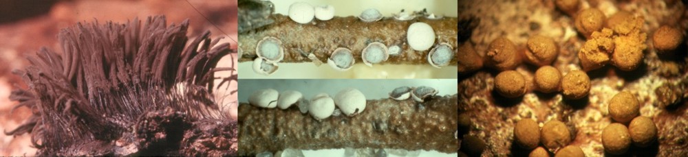
The final set of pictures illustrate Physarum bivalve, Reticularia lycoperdon and Fuligo septica. Physarum bivalve produces its spores in plasmodiocarps. These structures differ from sporangia in that they take the form of the original plasmodium and follow its branching pattern. Plasmodiocarps are usually net-like or made up of a series of elongated structures. There are some structures that are intermediate between plasmodiocarps and sporangia but usually the two types are easily recognized. Reticularia lycoperdon and Fuligo septica both produce their spores in aethalia. Aethalia are solid cushion-shaped structures bearing spores inside a firm to delicate crust. These can be large and rather irregular in form.
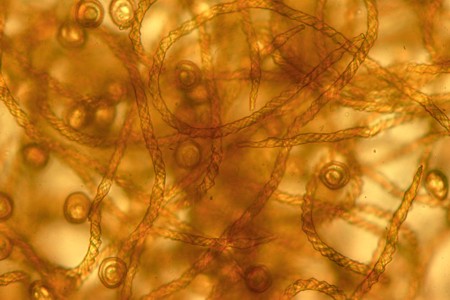
Slime moulds can also be interesting under the microscope. The wooly or hairy material emerging from the fruiting structures in some of the photographs is part of the capillitium, filamentous elements that respond to changes in humidity and propel the spores into the air. The capillitial threads often have striking spiral ridges that can aid in the identification of species. The capillitiam may be encusted with calcium carbonate, as may the outer shell or peridium of the fruiting structure. These lime deposits are also useful in indentification. The spores, which, depending upon the species, may be brightly coloured or dark brown, are usually nearly spherical and finely to coarsely roughened. These are often larger than most single-celled fungal spores and can be recognized readily when they are found in spore traps designed for air-borne fungi and pollen.
The relationship of slime moulds to fungi is remote. In fact, human beings are probably much more closely related to fungi than any of the slime moulds are. Most systematists today lump slime moulds in a "clade" called the Ramicristates. Included within the Ramicristates are a great number of organisms having amoeba or amoeba-like movement. This is an ancient and genetically diverse group, possibly comprising a dozen or more kingdoms of organisms.