Home >> Diversity and classification >> True fungi >> Dikarya >> Asexual Dikarya >> Hyphomycetes
HYPHOMYCETES
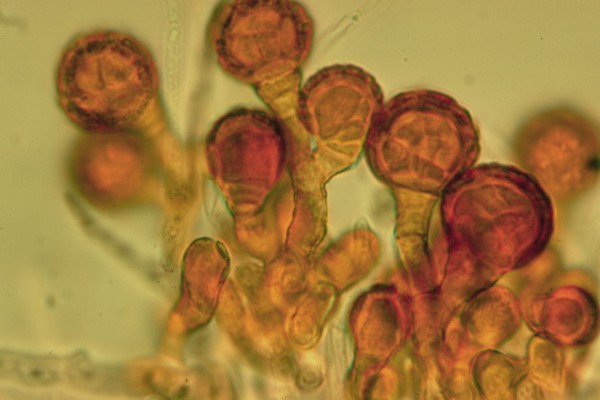
Hyphomycetes are asexual reproductive structures produced directly on their substrate without any kind of enclosing tissues. For example, Epicoccum nigrum, the fungus in the picture at left, grows directly on the surface of dead leaves and grass stems where it is exposed to the environment and can have its conidia (spores) carried away by air currents. Because of this its conidia are abundant in the air and are commonly found by people investigating airborne fungi. Mycologists working with fungal cultures in Petri dishes commonly get E. nigrum as an uninvited guest.
The most common function of hyphomycetes is reproduction and dispersal, although in some species the conidia may act as gametes or "spermatia" that can fertilize an incipient dikaryon. If this is so they are not likely to germinate and produce hyphae as happens in the great majority of hyphomycetes.
Hyphomycetes come in a staggering variety of forms. This immense diversity reflects the role these forms play in the dispersal of the fungus producing them. Each species grows in a particular habitat. When the nutrients in this habitat are exhausted the fungus must ensure that its offspring find their way to a similar source of nutrition. Getting their conidia to this new place, often a very small target, requires precise dispersal mechanisms.
Air dispersal
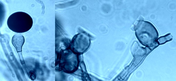
Epicoccum nigrum and Nigrospora sphaerica, are examples of fungi that actually propel their conidia into the air using mechanisms that force the conidium away from its subtending cell. This mechanisms is especially clear in N. sphaerica, the fungus pictured at right. In the left panel of the picture the large black conidium is still attached to its cell awaiting lift-off. In the right panel there are two cells that have already discharged their conidia; here we can clearly see the tube that directed water pressure up against the bottom of the conidium, shooting it away. We can also see the thin collar that originally held the conidium in place.
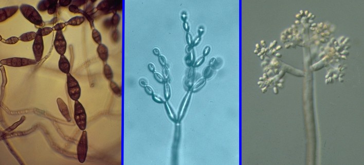
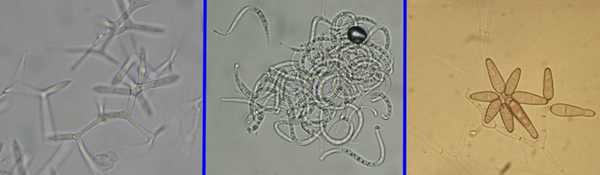
Most air-dispersed hyphomycetes do not have forcibly discharged conidia. Instead they have very loosely attached conidia that can be disloged by the slightest air current. Fungi of this type often have long hyphae that extend the conidia well away from the substrate, thus escaping the dead air (boundry layer) that surrounds them. At left are three species that occur commonly in samples of airbore fungi. These are, left to right, Alternaria alternata, Cladosporium cladosporioides and Botrytis cinerea. These three species are unrelated to one another yet often share the same habitats and have similar modes of dispersal.
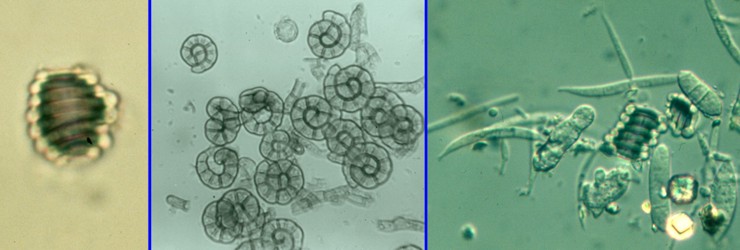
Water dispersal Many hyphomycetes are dispersed by water rather than air. Some of these species actually live out their lives in water while others only use water as a means of dispersing their conidia. The three photos above illustrate species using freshwater for their dispersal. At left is a species of Helicodendron, characterized by conidia forming a tight barrel-shaped helix. Such hyphomycetes are often called "aeroaquatic hyphomycetes" referring to the fact that the conidia, although dispersed by water, are able to trap air and float along on the surface. Such fungi often grow in habitats that are periodically flooded but are usually dry. When water levels rise the conidia are able to float away. The species of Helicoma in the middle panel also produces coiled conidia but these are spiral, not helical, and are thus produced in only two dimensions. These conidia are not as likely to trap air but will become attached to bubbles of foam. Spiral conidia often straighten out when they get wet, although that is probably not what happens with this species of Helicoma.
Conidia of aquatic hyphomycetes are easy to collect and observe under the microscope. They often collect and concentrate in the foam of running streams. To observe them, just collect the foam in a zippered plastic bag and bring them back for examination. The foam may gradually convert back to a liquid in the bag, but the spores will remain in abundance. Another way to observe these fungi, especially the aeroaquatic species, is to take a sample of forest litter that you suspect of being periodically wet and shake it up in a container of water. Then collect the very top layer of water with its floating debris and examine it under a microscope. Conidia will be abundant. The third panel in the photo above shows a sample taken from forest litter. An aeroaquatic Helicodendron species is clearly visible at the centre along with some other spores and unidentifiable debris. Also visible at upper middle is a narrow spore oriented horizontally and having an nearly vertical "arm", resembling a narrow canoe with a mast. This is a conidium of a Tricladium species, a good example of what are called "Ingoldian fungi", named in honour of Dr. C.T. Ingold, a pioneer in the study of aquatic fungi. Ingoldian fungi have conidia that are narrow, straight or loosely helical and often have radiating arms like the one in the picture. Aquatic hyphomycetes are also found in marine waters and can be observed in seafoam in much the same way they can in freshwater foam. The picture above illustrates three common marine species. At left is Varicosporina ramulosa, a species common on decaying seaweed and other debris in warmer climates. It does not occur in our cold New Brunswick waters but can be found a day's drive away on the south shore of Cape Cod, particularly in the summer. It is in every way a typical Ingoldian fungus. The middle panel shows a species that occurs commonly in New Brunswick. This is Sigmoidea marina, another Ingoldian fungus because of its lazily helical conidia. In the right panel is Asteromyces cruciatus, a species commonly encountered on decomposing seaweed in temperate climates but not yet found in New Brunswick. At first glance A. cruciatus would not appear to be a species having aquatic dispersal. However, the dark conidia
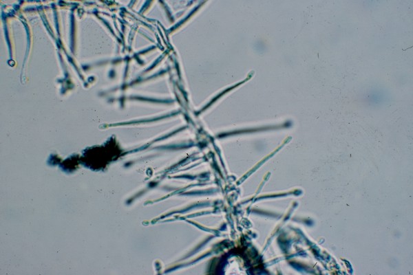
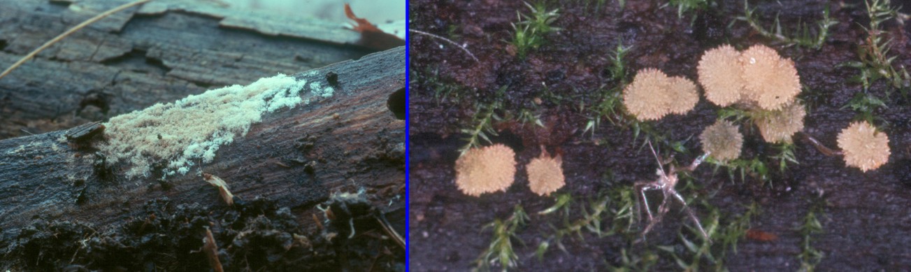
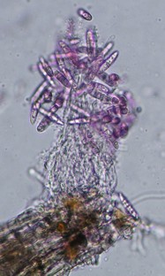
remain connected in star-shaped masses that function as typical Ingoldian fungi. Dispersal by insects and other small animals Insects, mites and other small invertebrate animals live among fungi and share many common difficulties. Such organisms, including fungi, have fairly precise nutritional and environmental requirements and may be separated from acceptable habitats by considerable distances. Reaching a precise habitat, especially one that is not large, may require more than just casting some spores to the wind. Insects, of course, can fly well and have eyes and sensitive chemical detectors to aid them get where they need to go. Fungi, lacking such abilities often solve the problem by hitching a ride on an insect. This solution to a difficult problem of dispersal is so successful that nearly all groups of fungi have made use of it. The hyphomycetes are among the most adept. The hyphomycete shown at right may be an excellent exploiter of insects for its dispersal. This fungus, a species of Acremonium produces its conidia at the tips of the small "spines" along the hyphal strand. The cells, called phialides, produce a continuous succession of conidia at their ends, and these conidia collect there in glistening drops of liquid, unfortunately not seen here because they flowed away when the microscope slide was prepared. Air currents do not dislodge the conidia nor are they likely to come in contact with water. Instead they stick to the bodies of insects and mites that are moving about in their habitat. The conidia are sticky because of the liquid they are borne in. Eventually the animal will move on to a new habitat and the fungus with be rubbed off and begin to grow there. Since many fungi live in enclosed places with few air currents to move them it is not surprising that many have taken the insect route. Organization of hyphomycetes The photo above presents two hyphomycetes growing on a log, Haplotrichum conspersum at left and Bactridium sp. at right. Note how different they are in their growth habit: H. conspersum forms an almost continuous carpet on the log, irregular in places but nevertheless all one surface of conidia. On the other hand, the Bactridium species has produced its conidia in discrete little cushions, well separated from one another. Species of Bactridium are said to produce their conidia in sporodochia, a term denoting a dense cluster of conidium-bearing hyphae or conidiophores. The conidiophores in Haplotrichum are said to be solitary because they do not form distinct clusters. In the two photos at left we see yet another variation. Here the conidiophores have united to form an elongated compound structure called a synnema (plural, synnemata). Synnemata are often associated with dispersal by insects. Pesotum ulmi, a synnematous form associated with the infamous Dutch elm disease, is well-known to transfer its spores to bark beetles, the vectors of the disease. The two photos represent the Graphium state of Pseudallescheria boydii at left and at right Arthrosporium albicans, an inhabitant of dead birch branches still covered by loose bark. Both occur in habitats frequented by insects and mites but not exposed to air currents. In Graphium species the conidia are borne in a droplet of liquid at the top of large dark synnemata. In A. albicans the conidia are borne on the upper part of the synnema but also occur along the sides. Identification of hyphomycetes Hyphomycetes are difficult to name. Identification requires a knowledge of developmental features and some skill with a microscope. To pursue this subject further you may wish to consult the Moulds pages at this website. Two criteria have been used traditionally for identifying hyphomycetes: colour and conidial septation. Colour is usually separated into two categories, light and dark. Darkly pigmented species are said to be "dematiaceous" while light or colourless ones are called "moniliaceous". Some annoying intermediates may be found. Conidial septation is usually divided into categories of one-celled, two-celled, more than two-celled but with transverse septa only and more than two-celled with both transverse and vertical septa. Although these criteria do not indicate genetic relatedness they do help in sorting things out.
Home >>
Diversity and classification >>
True fungi >>
Dikarya >>
Asexual Dikarya >>
Hyphomycetes