Home >> Diversity and classification >> True fungi >> Dikarya >> Ascomycota >> Pyrenomycetes >> Dothideomycetes >> Pleosporales and Hysteriales
THE PLEOSPORALES AND HYSTERIALES
The Pleosporales form a diverse group of fungi that are usually not difficult to recognize. Superfically they may resemble other pyrenomycetes, but under the microscope the prominant pseudoparaphyses and bitunicate asci distinguish them from most other groups. Things become more difficult within the Pleosporales because the large number of families and diversity of habitats encompass a great amount of variation. It is not practical here to go into the intricacies of classification within the Pleosporales, but we can as naturalists appreciate their ecological diversity.
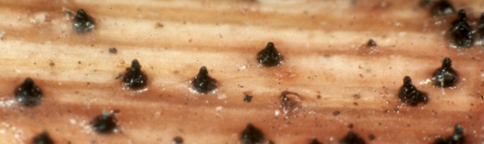
The photo above showing perithecia of Ophiobolus rubellus represents one habitat often dominated by Pleosporales, the overwintered stems of non-woody plants. The specimens in the photo were growing at the base of a still-upright dead stem of spotted touch-me-not (Impatiens capensis). This is a typical habitat for a large number of species that share a common type of life history. These fungi are mild to severe parasites of hebaceous plants showing some degree of host-specificity. Some, such as O. rubellus grow on dicots while others are found only on monocots, including grasses and sedges. They live on these plants throughout the growing season and initiate their perithecia at the beginning of the dormant season. In our region they can be collected in the autumn but will not contain mature asci and ascospores. They begin to mature at the onset of the new growing season, reaching maturity as the host plant is developing. In this way they are ready for ascospore discharge and colonization of the new host at precisely the right moment. The perithecia are usually on stems lying on the ground or near soil-level on those still standing. They are thus produced in relatively moist habitats and do not usually have features indicating a strong resistance to drying. Their peithecial walls are usually fairly thin and their asci are borne among wide hypha-like cellular pseudoparaphyses. If you wish to collect and observe these, look for them on dead plant stems occurring in moist places and at the time of year the new generation of the plants is appearing. This is a particularly good way to find members of the family Pleosporaceae.
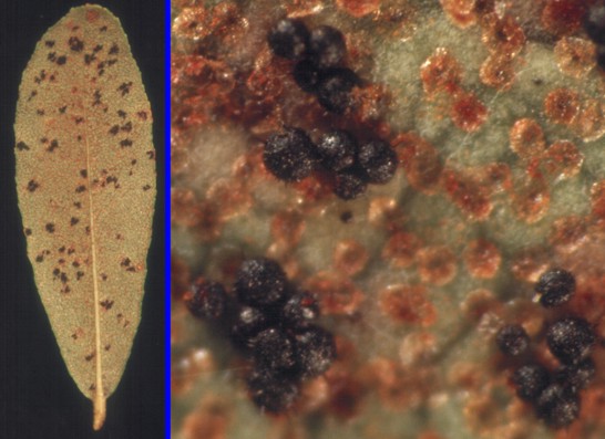
Other members of the Pleosporales also parasitize plants but specialize in hosts that are perennial. This is particularly common in the family Venturiaceae to which all our examples belong. Stigmatea pulchella, the fungus at left, is a good exaple of this kind of life history. The left panel is a photo of a leaf of Chamaedaphne calyculata, leather-leaf, bearing several colonies of S. pulchella, seen as black spots. The close-up in the right panel reveals that the black spots are actually clusters of tiny black perithecia. The orange-brown structures are peculiar scales that produced by the plant and are characteristic of leather-leaf.
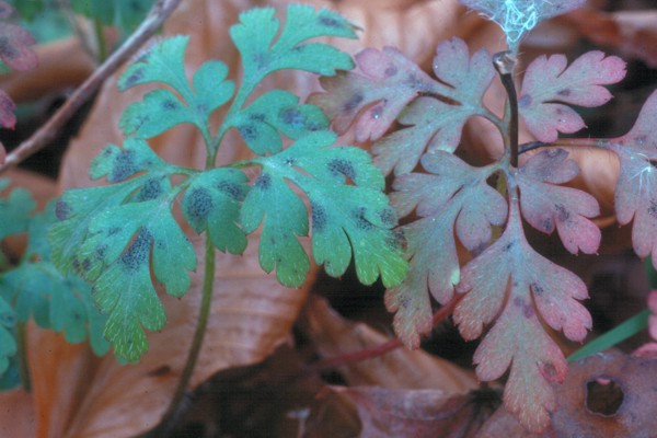
The life history of S. pulchella is similar to the one discussed above; the fungus matures in the spring and colonizes new leaves as they emerge. Members of this ecological group differ from the previous one in their production of perithecia on living leaves. Although these fungi grow in the relatively dry environment of a leaf surface they are active parasites that can tap directily into the host for moisture. As a result of this they do not have obvious means of combatting drought. Coleroa robertiani, a parasite of Herb Robert, has a similar life history. In the photo at right leaves of this little plant can be seen peppered with the small perithecia. They mature in the early spring and colonize the new leaves as they unfold.
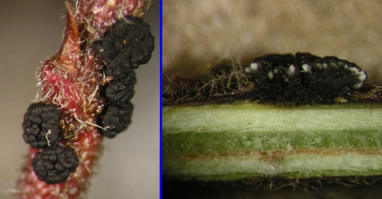
The pictures at left are of Gibbera vaccinicola a parasite of living blueberry stems. Although most blueberries are not large plants their stems may protrude above snow level, exposing them to the extreme desiccation of dry winter winds. In such situations it may be advantageous to have ascomata that are thick-walled and less easily dried. Gibbera vaccinicola produces large compound ascomata that can be easily seen with the unaided eye. The panel at the right of the picture shows a razor-blade section of an ascoma revealing the multiple clusters of asci inside. Each of the small white areas is one centrum composed of asci and pseudoparaphyses. Aside from its more massive structure G. vaccinicola has the typical life history of a plant parasite.
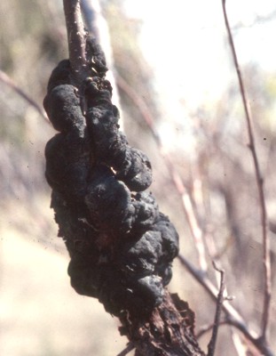
An even more massive ascostroma can be seen in Apiosporina morbosa at right. This species grows on wild cherries where it stimulates hypertrophic (abnormally fast) growth in the host plant, resulting in large swellings. The fungus produces its black ascostromata on the swellings, which may be several centimeters in length. Often the branches are killed at the outer end of the swelling. The disease, which can be severe at times, is called "black-knot disease of cherry". These ascostromata are home to hundreds of individual ascus clusters which in cross section look very much like those of Gibbera vaccinicola. The ascomata are often produced quite high in these small trees and would be exposed to even harsher conditions than those described above. Another species of Apiosporina, A. collinsii, produces its ascostromata on living leaves and twigs of serviceberry.
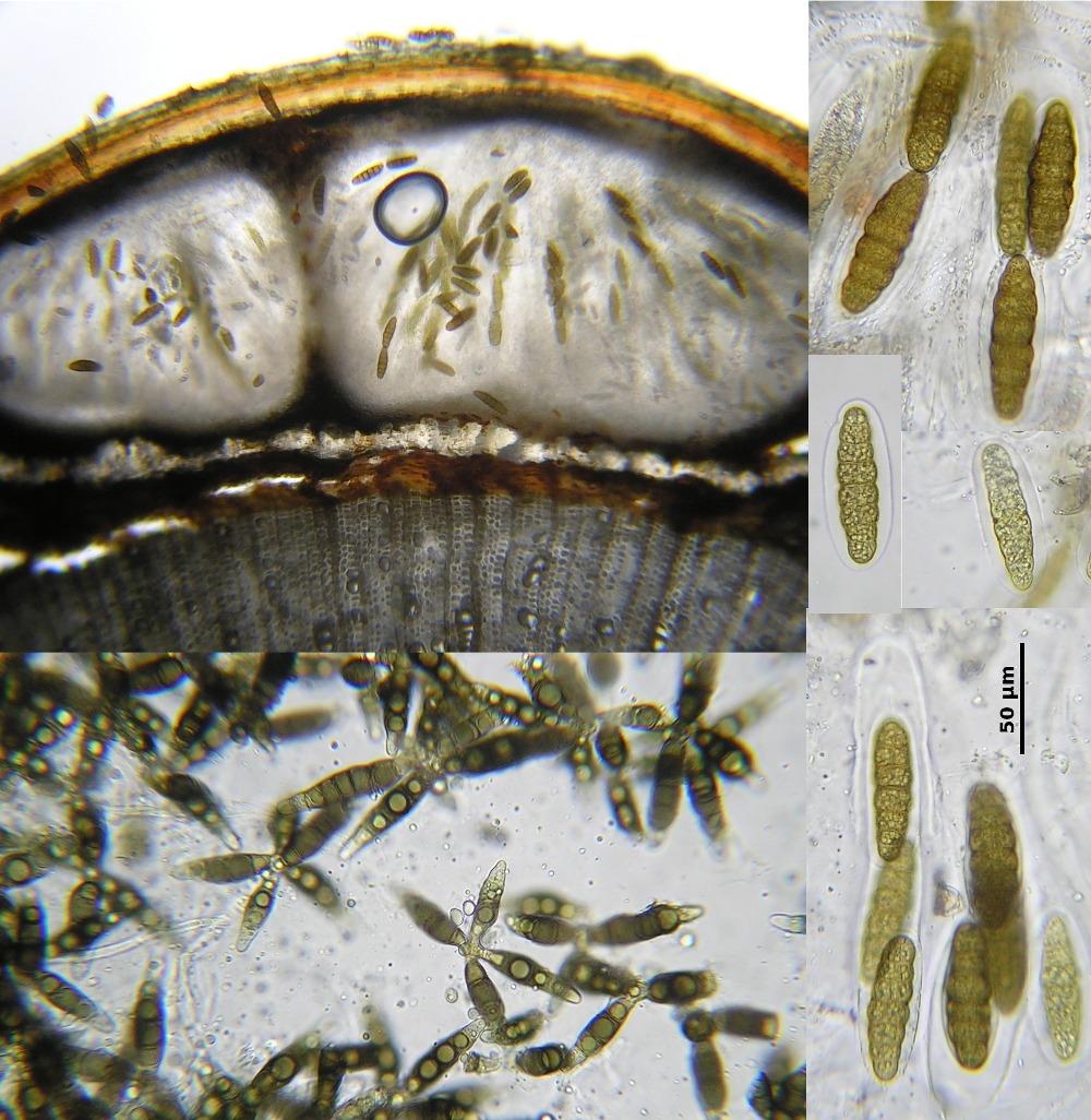
Pleomassaria siparia, figured at left, is a borderline parasite of birch trees. It belongs to the ecological group of fungi we call plant endophytes. Although its life history is not well-known, we think it probably enters small branches of the tree while they are still alive and healthy. Once established in the tree it probably remains inactive, absorbing only enough nutrients from the tree to sustain itself. When the branch dies the fungus begins to grow and reproduce. The photo at left shows two kinds of spores, its large ascospores, seen in the furthest-right panel, and its anamorph Prosthemium betulinum, a coelomycete.
If you have access to young birch trees you may be able locate P. siparia. Look for twigs 2 to 5 mm in diameter that have died recently. The fungus is inconspicuous and will be located most easily using a good hand lens or dissecting microscope. As the photograph shows, the perithecia are produced below the bark and reveal their presence by a convex swelling on the surface of the twig, broken slightly at its apex by the emerging ostioles. With good magnification you should be able to see the large ascospores adhering to the bark around the ostiole. The anamorph is similar in appearance under a lens, but of course differs greatly when viewed on a microscope slide.
Both ascospores and conidia of P. siparia are unusual, suggesting an unusual life history. The ascospores are surrounded by a thick gelatinous coating that probably accounts for their adherence around the ostiole. It is possible that these spores are ill-fated ones that failed to be discharged into the air or perhaps this behaviour is part of a successful dispersal method. If they are air-dispersed, the sticky coating would help them stick to branches of a living tree. The conidia are unusual in having several dark arm-like extensions radiating out from a central point of attachment, suggestive of the sort of pattern seen in spores of aquatic fungi. It is not known whether water has any part in the dispersal of the Prosthemium anamorph of P. siparia, but it is known that infections from some plant disease fungi can be spread by water running down branches.
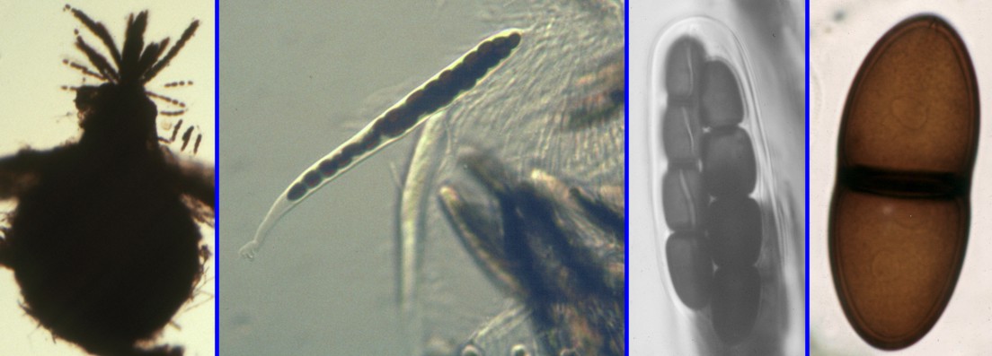
Not all Pleosporales are plant parasites. The picture above illustrates a group of species in the genera Delitschia and Preussia that are almost always found on dung of herbivorous animals. These coprophilous fungi are not a very large group within the Pleosporales yet are extremely common on dung. There are several genera of the Pleosporales on dung, especially members of the Sporormiaceae, Delitschiaceae and Trichodelitschiaceae, but species of Preussia are by far the most abundant and widespread. Some occur on dung in very dry habitats and may survive there for many months while others last only a few days. In the picture above the left panel shows a perithecium of P. dubia with a fascicle of asci that have been forced out of the ostiole. The next panel is a photograph of an ascus of P. dubia alongside material squeezed out from the perithecium. Note the "curtain" of pseudoparaphses in the upper right part of the picture. The third picture is a close-up shot of the ascospores of P. intermedia still inside the ascus. The ascospores are 4-celled, with each cell provided with a germination slit, a defining characteristic of Preussia species. At far right is an ascospore of a species of Delitschia. Species of Preussia have ascospores of four or more cells that often disarticulate into single-celled units. Species of Delitschia consistently have 2-celled ascospores that do not disarticulate.
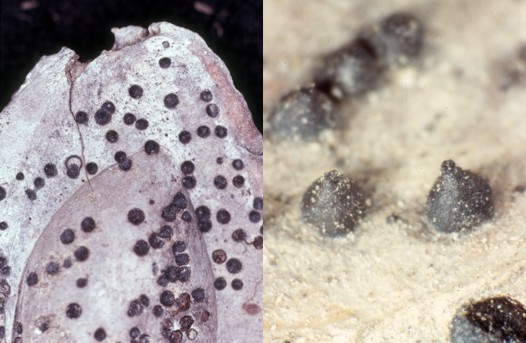
Caryospora putaminum, the fungus in the picture at left, is a representative of yet another ecological group of Pleosporales. These species grow on dead woody substances, often under very dry circumstances. They usually have very thick resistant perithecial walls and a dense trabiculate hamathecium embedded in a gel. They are apparently rather long-lived fungi that mature a few asci at a time and release their spores when an occasional rain moistens them. The hamathecium is able to remain dry for long periods of time and then rehydrate effectively because of the copious gel. The ascospores are often large, dark and thick-walled. These are very sturdy little fungi. Caryospora putaminum is restricted to the stones of peaches that remain on the ground after the fruit has decomposed. They are fairly easy to find in peach orchards and will usually bear mature asci. The left panel in the picture shows part of one peach pit covered in the perithecia of the fungus. the right panel is a close-up view of a few of these. Both the low and high magnifications reveal old, broken perithecia as well as intact ones. That particular collection contained perithecia in all stages of development, indicating that season plays a less important part in its life history than it does in that of the plant parasitic species.
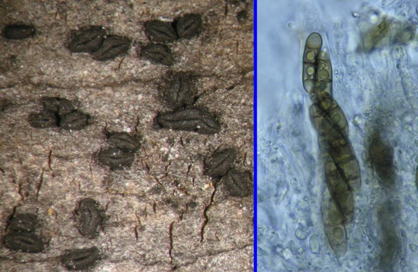
The plate at right presents yet another of the more sturdy members of the Pleosporales. This fungus, Platystomum compressum, grows on dead wood that is lying in a dry location. The particular collection used in the photo was made on maple firewood that had lain exposed for more than a year. Although the wood was periodically wetted by rain it was also subjected to drying. It differs somewhat from other members of the Pleosporales in having its ostiole flattened or elongated so that it opens as a slit rather than a round pore. Otherwise it is typical of this part of the order in having a trabeculate hamathecium and very thick resistant perithecial walls. The panel at right shows a group of asci, each with eight muriform (both vertical and horizontal septa) ascospores. In the upper left part of the picture you can see the thin pseudoparaphyses.
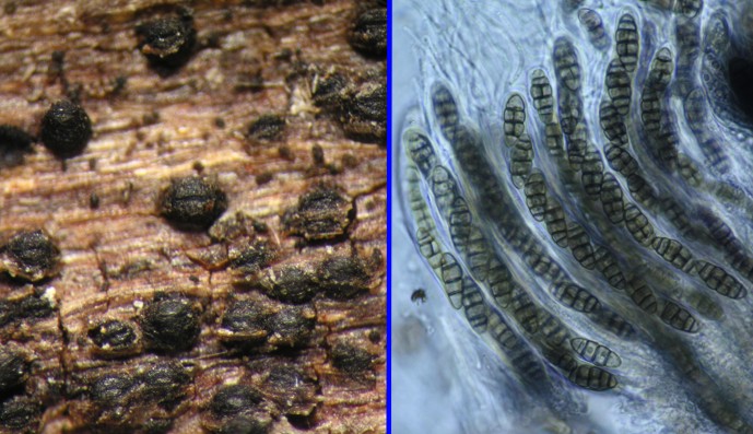
Platystomum compressum, with its slit-like ostiole and occurrence in demanding habitats, leads us conveniently from the Pleosporales to the Hysteriales. The organism in the plate at left is Hysterium pulicare a common inhabitant of the bark of hardwood trees. In fact, the picture here was taken from a specimen found on maple bark in the same woodpile as the previous fungus. The Hysteriales differ from the Pleosporales in producing hysterothecia instead of perithecia. Hysterothecia are elongated ascomata that open their entire length by a slit. Normally they are tightly closed, but when they become wet they will open to reveal the asci within. True to their dry habitat they have a trabiculate hamathecium and thick ascomatal walls. There are several genera and species in the Hysteriales, most of which are found on the bark of living and dead trees.