Home >> Diversity and classification >> True fungi >> Dikarya >> Asexual Dikarya >> Coelomycetes
COELOMYCETES

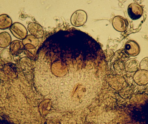
Coelomycetes are asexual Dikarya that produce their conidia inside some kind of enclosing structure. If this structure completely surrounds the conidia it is called a pycnidium; if the conidia are borne in a more open structure with a wall only around the lower part it is an acervulus. The large round structure in the picture at right is a pycnidium of the fungus Sphaerellopsis filum. This fungus is a parasite of rust fungi and occurs within their spore-bearing structures. In the picture you can see the urediniospores of the rust as numerous oval bodies surrounding the pycnidium of the Sphaerellopsis. The pycnidium here is very light coloured where it is sunken in the host tissues but dark on top. This is a common phenomenon among pycnidial fungi. Most pycnidia release their spores in a wet mass that is extruded out through the apical pore (ostiole). These often collect in large amounts and are either transported away by insects or will flow along with rain water.

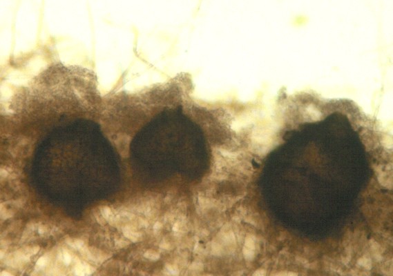
Growing as a parasite on rust fungi is a rather unusual place to find a coelomycete. More commonly these fungi grow on living and dead plants. Some can become a nuisance to humans when they grow where they are not wanted. The specimen of Phoma herbarum pictured at left was orginally found embedded and growing in silicone sealant used to waterproof a meat-packing establishment near Toronto. All of the sealant had to be removed and replaced. Species of Phoma regularly turn up on materials in indoor environments and may be responsible for some of the dark brown to black mould growth seen on painted surfaces subject to moisture, such trim around showers and bathtubs and on window frames and sills.

The pycnidia considered so far have been simple flask-shaped structures with a single internal cavity. However, some pycnidia and acervuli can be highly complex and be composed of several cavities. The picture above is an example of one of these. This fungus, Libertella betulina commonly occurs on recently-killed birch logs. The first signs that it is there will be the small orange tendrils arising through small breaks in the bark, as figured in the left panel. A closer examination (centre panel) reveals that these tendrils have oozed out like toothpaste from its tube. In the right panel the structures have been cut in half with a razor blade to reveal what is below the bark. Here we can see that there are several chambers each producing orange conidia in massive numbers. These are then extruded out through one or more ostioles.


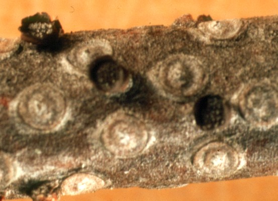
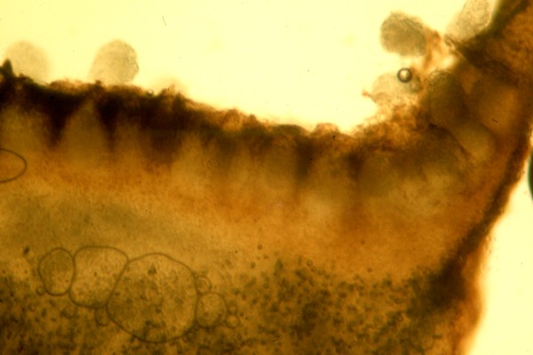
Bothrodiscus berenice, the species figured above, produces yet another sort of compound structure. Here the pycnidia are embedded together inside small goblet-shaped structures on the surface of balsam fir twigs. The picture at far left shows three of these "goblets" while the one to the right presents a cross section through the goblet to reveal the layer of pycnidia on its surface.