Home >> Diversity and classification >> Fungus-like organisms >> Oomycota
THE OOMYCOTA
Members of the Oomycota (pronounce both o's as you would in the common expression 'oh oh!') are all distinguished by their production of oogonia and oospores. Most lack septa in their hyphae and are therefore referred to as coenocytic. Unlike nearly all of the true fungi the Oomycota spend most of their lives in the diploid state as humans do. Only during the production of gametes (sex cells) do they produce any haploid cells. If you are not familiar with these terms you may wish to read through the page on fungal reproduction, especially the part on "gametangial meiosis".
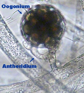
As in humans as well as plants and other animals, sexual reproduction in the Oomycota involves specialized reproductive structures where meiosis can occur and gametes formed. These structures (gametangia) are called antheridia and oogonia in the Oomycota. Antheridia are usually fairly small filamentous structures differing little from ordinary hyphae whereas oogonia are usually large and conspicuously swollen. The gametes within the antheridia are not produced as cells but only as nuclei containing the parental genome. On the other hand, the oogonia produce large cellular structures called oospheres, each containing a haploid nucleus. During reproduction the antheridium fuses with the wall of the oogonium and perforates it, allowing the antheridial nuclei to enter first the oogonium and then the oospheres themselves. The nuclei then fuse within the oospheres to produce a diploid. The fertilized oosphere subsequently develop a thick wall, becoming mature oospores. Oospores are often produced at the end of the growing season and must be resistant enough to survive until conditions are suitable for new growth. In the picture at left the antheridium is arising from the same hypha supporting the oogonium, suggesting that this is a case of self-fertilization. However, in many species fertilization is between gametangia of separate individuals. It is probably also likely that an individual will fuse with either its own gametangia or those of another colony, whichever presents itself first.
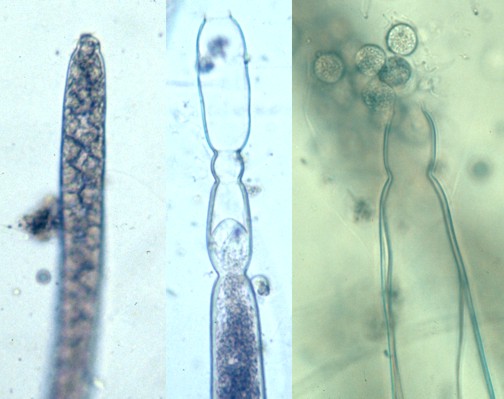
Asexual reproduction is highly varied among the Oomycota and is, in fact, the most convenient means for identifying them. The fundamental type is a simple sporangium resembling the end of a hypha containing numerous diploid asexual spores. The spores are often motile (zoospores), swimming by means of paired of flagella. The newly discharges spores swim vgourously away when they are released from the sporangium in many species, but in some they may form non-motile cysts immediately , such as those in the photograph at right. The three sporangia in that picture were all produced by a species of Achlya and show three stages of sporangium-formation and spore release. The sporangium at far left is nearly mature and has clearly-dfined spores. The one in the middle has discharged its spores and is starting to form a new sporangium inside the walls of the old one by a process called percurrant proliferation. The sporangium at right has discharged its spores and these can be seen near the opening.
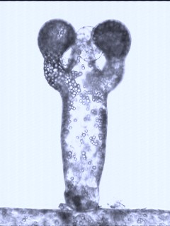
With their primarily diploid life cycles and large oogonia it is not surprising that the Oomycota might be rather distantly related to the true fungi. Most biologists now classify them with the Kingdom Straminopila, a fairly large group containing mostly photosynthetic organisms such as diatoms and yellow-green algae. The structure in the photo at left, looking like a triumphant boxer, is a reproductive element of Vaucheria cruciata a yellow-green alga common in freshwater ponds. Species of Vaucheria are very similar to members of the Oomycota except that they are photosynthetic. If the photo were not in black and white the alga would have a nice yellow green colour. Unlike members of the Oomycota Vaucheria cruciata produces two or three oogonia per structure with the antheridium in the middle. Thus the "boxing gloves" are the oogonia. Other species of Vaucheria have less elaborate reproductive structures and more closely resemble members of the oomycota.
The Oomycota is not a large group but is quite diverse, both in the appearance and the activities of its members. Although it has been divided up into as many as 30 families we generally view these organisms as being of two types, the water moulds and the plant parasites. This division certainly does not reflect the true genetic diversity of the Oomycota nor does it preclude several intermediates but it does help us to sort out two major trends in their natural history.
THE WATER MOULDS
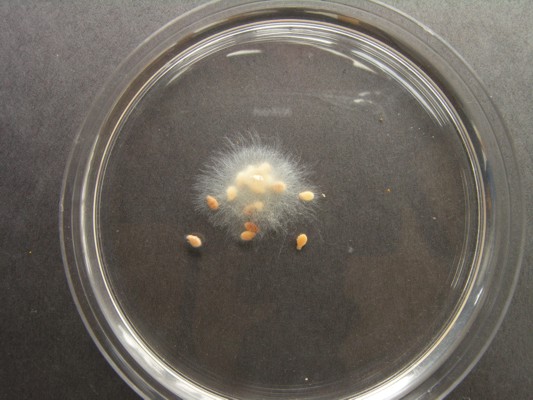
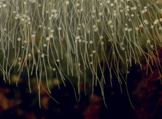
As their name implies, the water moulds are mostly aquatic. Any body of water, fresh or salt, contains them in abundance. These are easy organisms to grow at home and they are large enough to be seen easily with a low-power microscope. The pictures above show what you can do. A small amount of pond water is placed in a covered dish and sprinked with a few toasted sesame seeds. Toasting the seeds in a frying pan serves two purposes, it sterilizes them and it breaks the seed coat, allowing oils to escape. Within three or four days water moulds will begin to grow on the seeds. One more step was taken for the picture: here a colonized seed from the original sample was transferred to a new covered dish containing sterilized (boiled) water and a few fresh seeds. The culture may not be entirely free of other organisms, but there is probably only one water mould there. The picture above right right is just a more greatly magnified section of the colony. Here you can see white oogonia produced abundantly along the hyphae. Try this yourself; you will be amazed at the variety of water moulds you can find in a few different ponds, streams and ditches. You may also become fascinated with the other forms of aquatic life developing in your dishes.
Water moulds do not just live on seeds. Many are all-purpose scavengers, seeking out any form of organic material at hand. Some appear to be somewhat more carnivorous and are attracted to dead animal materials. The experiment described in the previous paragraph can be carried out equally well with dead flies. Others will colonize pieces of fruit, potato, and egg yolk. Often baiting a pond water sample with several different materials will yield a greater variety of species.
Some water moulds are parasitic, either on one another, algae, plants or on living animals. Some species of Saprolegnia can cause an epidemic in aquaria, killing all of the fish. Pet stores often carry substances that will act against Saprolegnia infestations. Many microscopic animals fall prey to water moulds as well
The division between water moulds and the plant parasitic species is not always completely clear. For example the genus Pythium. a common water mould in ponds and ditches, can also exist in moist soils and attack plants. Diseases of plants caused by Pythium species can cause serious problems in nurseries. The most serious of these diseases are the ones called damping off, discussed further in the section dealing with plant parasites.
THE PLANT PARASITES
The members of the Oomycota living as plant parasites may have some characteristics in common with the water moulds but are highly specialized for their mode of life. Many occur in situations where dispersal by water is impossible and lack motile spores. Sporangia are often deciduous and carried by wind to new plant hosts. Although most produce oogonia and oospores, these are often much smaller and contain only a single oospore.
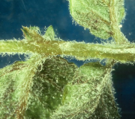
The picture at right shows part of a potato plant infected with a downy mildew caused by Phytophthora infestans. Note that the whole plant seemed to be covered with a white fuzzy growth. Downy mildews are among the most important of plant diseases caused by members of the oomycota. The progress of the disease is rapid, with the time between the first symptoms and total crop destruction being only a few days. Once a farmer discovers he has an outbreak of downy mildew it may be too late. Downy mildews have been the cause of immense epidemics in grapes in France, particularly during the nineteenth century, tobacco in Ontario in the 1990's and of potatoes in Ireland in the 1840's. The Irish potato blight was a particularly devistating event leading to starvation and mass emigration. There are many web sites discussing the Irish potato famine in detail so it will not be dealt with further here other than to point out that Saint John, the home city of this web page, sometimes called the Irish City, would have had a very different history were it not for this calamity.
The downiness of downy mildews is caused by its sporangia and the hyphae bearing them. These extend out from the surface of infected parts of the plant, gaining access to air currents. The significance of these sporangia and their relationship to the types of sporangia illustrated further up this page bears some examination.
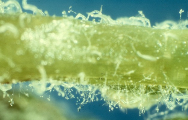
 Species of Phytophthora have deciduous sporangia that produce motile spores if they come to rest in an appropriately moist location. Some other species of Oomycota, in genera other than Phytophthora, also have deciduous sporangia, but these sporangia germinate by the production of hyphae and not zoospores. Thus we see in this group a trend toward less dependence on water for reproduction. The photo at left shows a close-up of a potato leaf infected with P. infestans. Here is is possible to see the individual sporangia extended away from the plant. These of course produce motile spores so we can expect that P. infestans would thrive during wet weather, which was exactly what was happenning when the Irish potato crop was devastated.
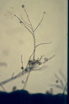
Finally at right is one further step in magnification where we can see additional details of the sporangia and supporting hyphae. Some of the egg-shaped sporangia remain attached while others have broken loose and are free to drift away.
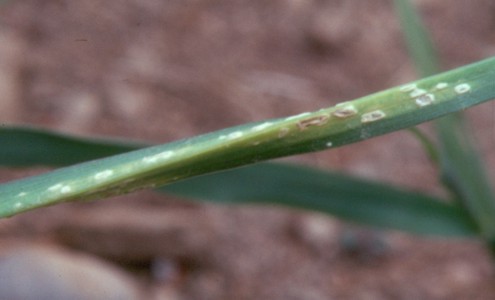
The white rusts, another group of plant parasitic Oomycota, are shown at left. These organisms produce chains of sporangia in dense groups below the epidermis of their host plant. At maturity the mass of sporangia pushes up, rupturing the epidermis and exposing the sporangia to air currents. The sporangia behave as though they were spores, germinating to produce hyphae. The species shown here is Albugo tragopogonis forming white blisters (sori) on the living leaves of goatsbeard (Tragopogon pratense). They are called white rusts because of their resemblence to the true rust fungi