Main page <> Index of descriptions <> Previous description <> Trichosporon <> Next description
Trichosporon
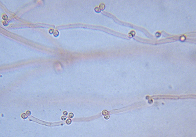
Species of Trichosporon are often difficult to recognize. The colonies are usually rather flat and a pale pinkish buff or yellowish colour. At first hyphal, the older parts of the colony become disarticulated so that often no hyphae are seen at all. Reproduction is by production of thallic conidia or by holoblastic conidia produced by peg-like processes on the hyphae. Some species may produce endoconidia as well.
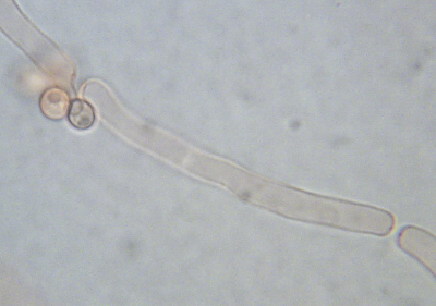
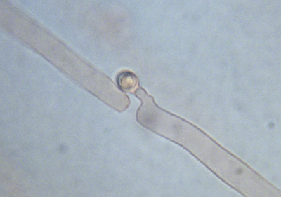
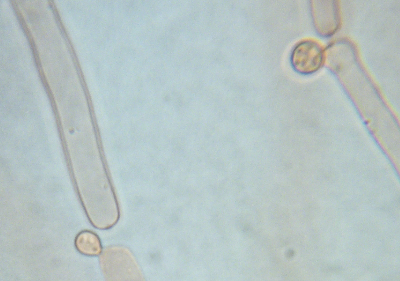
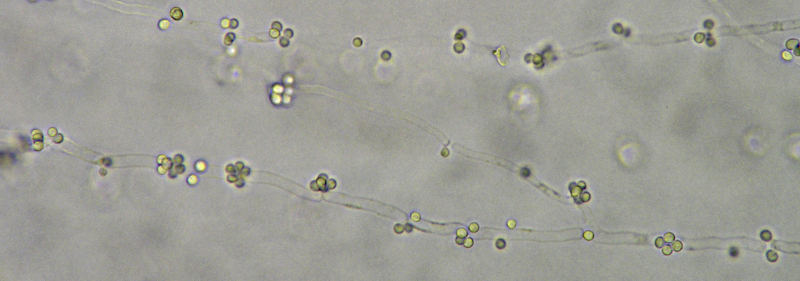
The pictures above illustrate some of the features of this rather enigmatic genus. The first four illustrate an isolate of T. laibachii from horse dung collected by Mr. Joop van der Lee in The Netherlands and identified by Dr. Allison Walker using DNA sequences. These all depict young parts of the colony, but even here many of the hyphae have become separated from one another. It's difficult to say whether or not these separated cells represent thallic conidia, but they certainly would be capable of growth to form a new colony. If you look very closely you will notice that the tips of the separated hyphae end with a small dark dot. Although obscure here, these little dots are dolipores, highly complex structures found only in basidiomycetes, which is what species of Trichosporon are. Dolipores function as minute valves controlling the flow of cytoplasm (cell contents) along the length of the hypha.
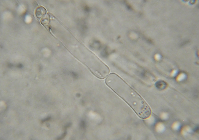
The long picture is of an unidentified species of Trichosporon quite similar in appearance to T. laibachii. It was isolated from decomposing seaweed (wrack) found on a beach beside the Bay of Fundy. Notice how here, again, the conidia are produced in clusters at the septa.
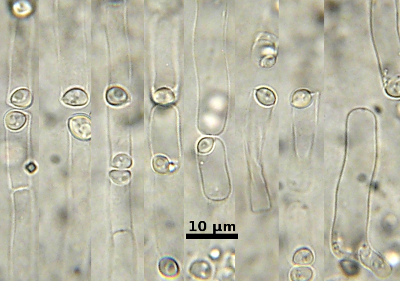
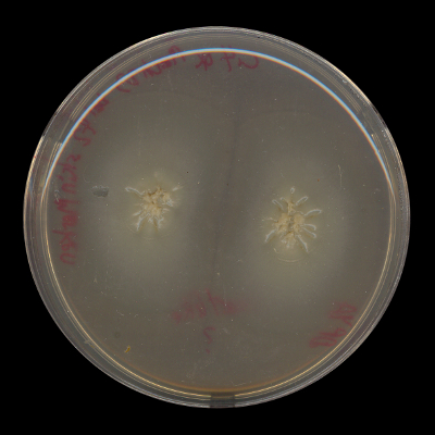
The remaining three pictures are of an isolate of T. dulcitum obtained by Karen Vanderwolf from the surface of a hibernating bat in New Brunswick. This isolate differes from T. laibachii in having its conidia borne inside the hyphae. These are rather unusual in appearing to be half inside the cell and half outside in some cases. However, like T. laibachii this isolate becomes almost totally converted to spores at the centre of the colony. The picture of T. dulcitum growing in a Petri dish is typical of many species of Trichosporon, showing the rather featureless, moist, yellowish colonies.
Identifying species of Trichosporon is challenging. The two named isolates used in the above illustrations had their identity confirmed by molecular genetic techniques, kindly carried out by Dr. Allison Walker for T. laibachii and Dr. André Lachance for T. dulcitum. Sugita's (Sugita, 2011) key to species employs the traditional methods used by yeast taxonomists, stressing nutritional and other physiological responses in broth culture. The species are remarkably similar to one another under the microscope, so that the molecular approach may be the only reliable way to sort them out.
Classification: Trichosporonaceae (Tremellales). Holomorph: Unknown. Ref: Sugita (2011).