Main page <> Index of descriptions <> Previous description <> Pestalotiopsis <> Next description
Pestalotiopsis
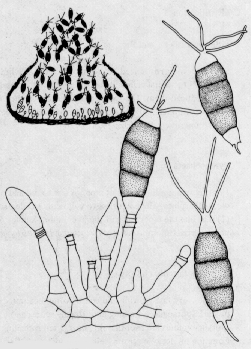

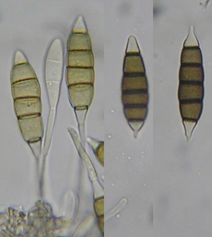
Conidiophores (annellides) produced within compact fruiting structures (aecervuli or pycnidia). Spores (conidia) 4- to 5-celled, with the two or three central cells dark brown, and with two or more apical appendages or hairs; collecting in a wet mass outside the aecervulus.
Pestalotiopsis is just one of a complex group of fungi. For example the photo at right is of Seiridium abietinum, a species found on dead branches of balsam fir in Atlantic Canada. It differs from species of Pestalotiopsis in having conidia with a single apical appendage rather than several. The following key may be some help in distinguishing these difficult genera. However, anyone seriously interested in identifying a member of this group should consult the monumental work on coelomycetes with appendaged conidia by T. R. Nag Raj (Nag Raj, 1993).
A SIMPLIFIED KEY TO PESTALOTIOPSIS AND RELATED GENERA
1. Conidia with a single apical branched or unbranched appendage 2
1. Conidia with appendages arising at more than one point on the apical cell 10
2. (1) Basal appendages always lacking 3
2. Basal appendage present in some of the spores 6
3. (2) Apical appendage laterally branched, comb-like
Labridella
3. Apical appendage unbranched or with branches regularly produced on more than one side 4
4. (3) Apical cell with a single unbranched appendage produced at a nearly right angle to the axis of the spore
Bleptosporium
4. Apical cell with appendage(s) branched or arising at several points 5
5. (4) Branches of the apical cell arising at one point or nearly so
Hyalotiella
5. Branches of apical appendage arising at several points
Truncatella
6. (2) Conidia without appendages commonly occuring among those with appendages
Seimatosporium
6. Conidia consistently appendaged 7
7. (6) Basal appendage arising from the basal septum of the conidium, often visible inside the conidiogenous cell before the conidium has broken off 8
7. Basal appendage arising from the lateral wall of the basal cell, usually visible alongside the conidiogenous cell before the conidium has broken off 9
8. (7) Conidia with four or five cells, euseptate, with normally thickened or relatively thin septa
Monochaetia
8. Conidia with six cells, disto-septate, with inner walls greatly thickened and often with conspicuous septal pores
Seiridium
9. (8) Apical appendage lateral, branched
Doliomyces
9. Apical appendage unbranched
Sarcostroma
10. (1) Conidia disto-septate, with inner walls greatly thickened and often with conspicuous septal pores
Pestalotia
10. Conidia euseptate,with normally thickened or thin septa 11
11. (10) Median (coloured) cells of the conidia thin, smooth-walled and pale, occasionally nearly colourless; apical appendages consist of one obliquely bent terminal appendage giving the spore a hummingbird-like appearance and one or more lateral appendages borne on the convex side of the apical cell
Zetiasplozna
11. Median cells of the conidia thick-walled, dark and sometimes roughened; apical appendages less regular in configuration
Pestalotiopsis
Parasitic or endophytic on living leaves and twigs but are often isolated from dead plant matter and even soil.
Classification: Pestalotiopsis species, and those of related genera, are anamorphs of members of the ascomycete family Amphisphaeriaceae (Sordariomycetidae). Members of the Amphisphaeriaceae and their anamorphs include Amphisphaeria (Bleptosporium), Blogiascospora (Seiridium), Broomella (Pestalotia and Truncatella), Discostroma (Seimatosporium), Ellurema (Hyalotiopsis), Griphosphaerioma (Labridella), Lepteutypa (Seiridium), Neobroomella (Pestalotia), and Pestalosphaeria (Pestalotiopsis). Doliomyces, Monochaetia, Sarcostroma and Zetiasplozna have not yet been found associated with a holomorph. Ref: Guba 1961, Kang, et al 1999, Nag Raj, 1993.