Home >> Diversity and classification >> True fungi >> Dikarya >> Ascomycota >> Pyrenomycetes >> Sordariomycetes >> Sordariomycetidae
THE SORDARIOMYCETIDAE
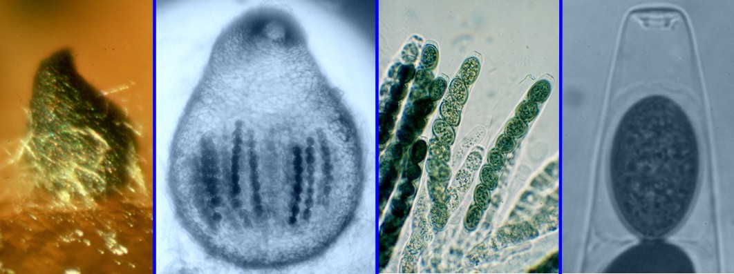
The genus Sordaria can serve us as a model of the subclass as a whole. While having most of the fundamental features of the group it is rather "stripped down" and lacks some of the complexity of other members. The illustration above shows several features of Sordaria; in the left panel a single perithecium sits on its substrates (agar in this case) where it is attached by colourless hyphae. It is pear-shaped and has a narrow neck leading up to the ostiole, the hole through which the mature ascospores can escape. In the next panel is another perithecium, but this one has been back-lit to show the asci and spores inside. Note also the prominant ostiole and the stone-wall appearance of the peridial cells. The next panel shows a group of asci, each containing eight dark green to nearly black single-celled ascospores. The right panel is a close-up of one ascus showing the conspicuous apical pore found in most members of Sodariomycetidae.
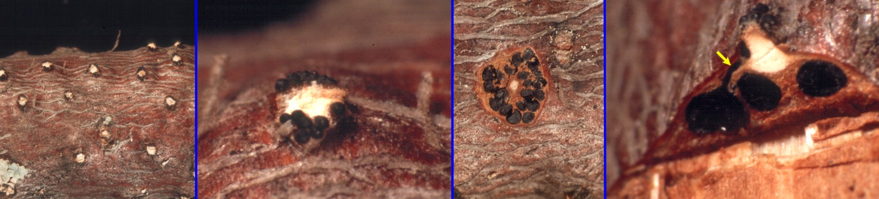
A more complex example of the Sordariomycetidae can be seen in Melanconis marginalis figured above. This species is a common inhabitant of recently dead alder branches where it can be seen as numerous small blisters (left panel). The next panel shows a close-up of one of the blisters, where the tips of the individual perithecia can be seen. Cutting across one of the blisters, as in the third panel, shows the perithecia seated in a circle around the central stromatic core. The last panel illustrates a rough cross-section through a blister to show the perithecia, again seated around the central core. The two perithecia at right were not cut exactly through their centres so you cannot see the necks, but the leftmost one is intact, showing the long neck, at the yellow arrow, emerging at the perphery of the core. Thus what we see in the second panel are the tips of the necks of the perithecia seated beneath the bark. The Austrian mycologists Dr. Walter Jaklitsch and Dr. Hermann Voglmayr have recently published a beautifully illustrated account of this and other species of Melanconis (Mycokeys 63: 69-117. 2020). The pattern of perithecia arranged in a circle with convergent ostioles is termed "valsoid" because it is most well-known in the genus Valsa. Valsoid perithecial groups are very common in the Sordariomycetidae.
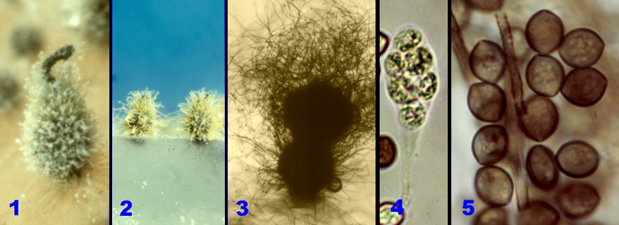
The genus Chaetomium, figured above, displays yet another variation on the Sordaria theme. This is a group that often grows in unexposed situations that do not necessarily favour forcible discharge of ascospores. Instead of shooting its ascospores out through the ostiole as other Sordariomycetidae do species of Chaetomium release them inside the perithecium where they gradually emerge in a large mass or in a tendril. The perithecia are covered in hairs, particularly on the upper parts, and the spores often become concentrated among them. It is not known how these particular characteristics function in the dispersal of the ascospores but they are obviously very effective; species of Chaetomium are widespread throughout the world, abundant and very diverse. Several aspects of Chaetomium are illustrated above: 1) a perithecium of C. subspirale showing a tendril of ascospores emerging from the ostiole in the manner of toothpaste from a tube, 2) two perithecia of C. cochlioides growing in a Petri plate, 3) a perithecium of C. globosum, perhaps our most common species, showing a nearly spherical mass of ascospores seated among the hairs at the ostiole, 4) an ascus of C. globosum showing the thin walls and lack of apical pore typical of asci that do not actively shoot their ascospores, and 5) ascospores of C. globosum, some with apical pores visible.