Fleshy Fungi of New Brunswick >> Galerina cerina
A Focus on Species >> Galerina cerina
Galerina cerina (Fr.) Kühner
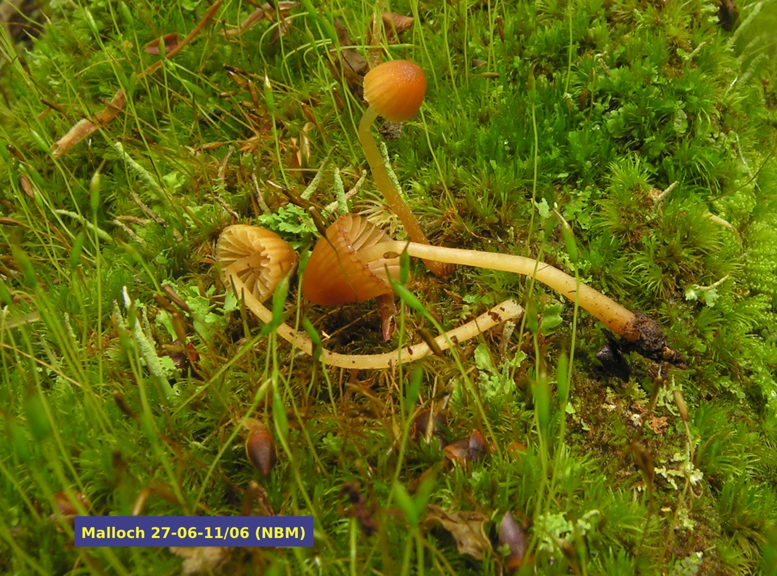
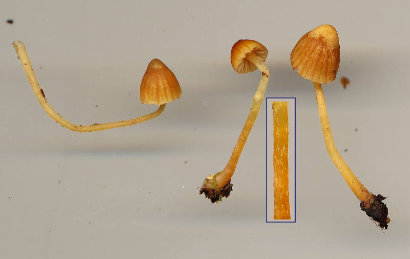 Galerina cerina is a small and easily overlooked mushroom. It is remarkably similar to several other species of Galerina, and is, in fact, only identified with certainty using a microscope. The ones shown on this and the main page were barely 35 mm tall and were found growing among mosses on an old sugar maple log in the Caledonia Gorge Protected Natural Area near Riverside Albert, New Brunswick. A comparison of the two illustrations is interesting: the three mushrooms were first photographed in the field and were then, several hours later, copied on a flatbed scanner. Note that the stipes are less transluscent in the older specimens and the one in the middle shows some white threads left over from the universal veil, seen better in the magnified insert. The caps have faded at the centre, a condition mycologists describe as hygrophanous.
Most species of Galerina grow in association with mosses, although the nature of this association is still poorly understood. Dr. Scott Redhead demonstrated that G. paludosa is actually parasitic on species of Sphagnum (2). Galerina cerina is usually found among species of the moss genera Dicranum and Polytrichum, although other genera, even Sphagnum spp., may be present. It is most common in spring and early summer when other mushrooms may be hard to find. The collection illustrated above was collected on June 27, 2011. Knudsen and Vesterholt (1) report it to occur from late spring to autumn in northern Europe.
 As pointed out previously, G. cerina cannot be identified confidently without a microscope. However, there are certain features that help to narrow the field down to some extent. Although G. cerina is not one of the species of Galerina having a really bulbous stipe, the stipe is nevertheless swollen or club-shaped at the base. The stipe itself is pale in colour when fresh and marked by white fibrils. These fibrils are the remains of a universal veil that enclosed the entire mushroom before it had elongated and expanded. They might also be visible at the margin of very young caps, although the ones in the photo do not show this. The closely related species G. calyptrospora has a more pronounced veil that can be easily seen at the margin of the cap when young. The gills of G. cerina are fairly pale and are adnexed, meaning that they turn up where they meet the stipe. The odour and taste of the flesh is rather nondescript, in contrast to the similar G. calyptrata which is farinaceous (mealy or like cucumber). It is necessary to point out that tasting species of Galerina carries some risk. Some species, such as G. marginata, are highly poisonous, even fatally so. The safe way to assess the farinaceous characteristic is to crush a cap between your thumb and forefinger and then smell it. Tasting is a slightly more sensitive test allowing the use of only half a cap, but you must be sure to rinse out your mouth after doing so and not swallow any of the mushroom.
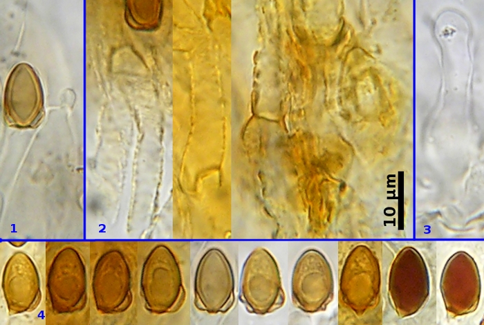
The most reliable way to identify G. cerina is with a microscope. Its most recognizable microscopic feature is the calyptra on its basidiospores. Figure 4 shows several basidiospores, the first four from the left are in profile; the next four are in face view, all mounted in 5% KOH. The two spores at right, seen in profile, were photographed in Melzer's solution to demonstrate the dark red dextrinoid reaction characteristic of most species of Galerina. Most of the spores in Figure 4 show a peculiar ballooning of the wall in the lower part. This swelling is called a calyptra and is the outer spore wall that pulls away from the spore is a very characteristic and regular way. Galerina is a fairly large genus of mushrooms with perhaps more than 200 species (3), yet no more than 20 of these species, such as G. hypnorum and G. sphagnicola, have calyptrate basidiospores. Figures 1, 2 and 3 show other features of G. cerina. Aside from showing yet another calyptrate spore, Figure 1 also shows a hypha with a clamp connection, a feature missing in some species of Galerina. The middle panel of Figure 2 also shows a hypha with a clamp connection. The main purpose of Figure 2 is to demonstrate the strongly encrusted hyphae that make up the surface of the cap. Figure 3 shows a cheilocystidium. These structures line the edges of the lamellae and are often useful for identification. Cheilocystidia of G. cerina are thin-walled and shaped somewhat like little bowling pins.
USEFUL REFERENCES
1. Knudsen, H and Vesterholt, J. 2008. Funga Nordica. Nordsvamp, Copenhagen. 965 pages + DVD.
2. Redhead, S.A. 1981. Parasitsim of bryophytes by agarics. Can. J. Bot. 59: 63-67.
3. Smith, A.H. and Singer, R. 1964. A monograph of the genus Galerina Earle. Hafner Publ. Co., New York. 384 pages.
Photo: D. Malloch