Fleshy Fungi of New Brunswick >>
Galerina sphagnicola
Galerina sphagnicola (G.F. Atk.) A.H. Sm. & Singer
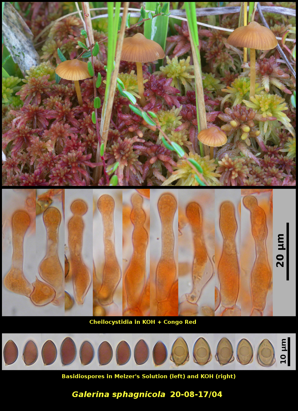
Gregarious (several) amongst sphagna on a floating lakeside bog, most closely associated with Larix laricina and Picea mariana, Spednic Lake Protected Natural Area, New Brunswick (20-08-17/04)
Basidiospores medium brown in spore print, almond-shaped, smooth, conspicuously calyptrate in KOH mounts, strongly dextrinoid, 8.1-9.7 X 5.0-5.8 μm, D/d = 1.50-1.76 (average[23]: 8.8 X 5.3 μm, D/d = 1.65). Cheilocystidia forming a continuous sterile margin, irregular in form, mostly with a swollen venter, narrow neck and swollen apex, occasionally moniliform.
Fruiting bodies of Galerina sphagnicola are easily confused with those of Galerina sphagnorum. Both occur with Sphagnum mosses, have long stipes and lack conspicuous veil material. They cannot be distinguished with certainty without microscopic examination of the basidiospores. Mounted in KOH, many of the basidiospores reveal characteristic "ears" caused by the loosening of the outermost wall layer. Spores of this type are said to be calyptrate and can be observed in several species of Galerina, such as G. cerina. The basidiospores in the plate here were photographed partly in Melzer's Soltion and partly in KOH. Those in KOH clearly show the calyptra, while those in Melzer's Solution mostly do not. The distinctions between the two species is discussed in more detail under G. sphagnorum.
You may have noted an apparent size difference in the basidiospores photographed in the two mounting media. The ones photographed in Melzer's Solution were taken from a spore print and can be assumed to be mature. Those mounted in KOH were taken from a piece of lamella from the same fruiting body used for the spore print. The size difference is real. The basidiospores from KOH measured 8.8-10.0 X 5.5-6.2 μm, while those from Melzer's Solution yielded the measurements given in the desciption above. It is possible that this difference in size is due to the different mounting media, but we have also commonly found that basidiospores measured from lamellae are larger on average than those taken from a spore print. This may be because the lamellae bear mature along with immature basidiospores, and we consciously avoid measuring the smaller ones we assume to be immature. Or maybe spores just shrink a bit after they have been discharged.
The basidiospore dimensions given by Smith and Singer in their world monograph of Galerina (Hafner Publ. Co., NY. 1964) are 9-11.2 X 6-7.3 μm, larger than the those of 20-08-17/04, regardless of how they were measured. They also mention a collection made by Dr. Howard Bigelow in Massachusetts that has monilioid (like a chain of beads) cheilocystidia, perhaps similar to our collection. These variations in spore size and cheilocystidium shape might be interesting to investigate.
Photograph: D. Malloch (20-08-17/04).