MUSHROOM COLOURS
Colours
Colour, one of the most important characteristics used in the identification of mushrooms, is likely to change drastically when the mushroom is dried. When we record notes on our fresh collections we must necessarily pay close attention to colour. An excellent way to do this is to take photographs of the specimen even before it is removed from its substrate. Some colours fade quickly and may change even before the mushroom is carried back to the work area. Immediately upon return it is helpful to photograph the mushroom under controlled light or even scan it with a flatbed scanner. Colour rendition of scanners is remarkably good. The photos and scans can be labeled with the number of the collection for later use.
Although photographs and scans are extremely useful when identifying mushrooms they may not be available to the identifier. When this is the case it is necessary to rely on the descriptive notes.
 For example, how would you describe the colours on the pileus of Hebeloma velutipes at right? Not easily, you might say. It's colours seems to be a complex mixtures of brown except for the grey margin. Of course "brown" and "grey" are not good enough because there are innumerable shades of both. You need to be more specific. At its very centre there is a small area of light orange. Outside that area is a region of a pale yellow-brown to pale olive-brown. Further out the colour is a very pale yellow brown with fewer olive tints, and then finally the margin is pale grey. This brief description is still imprecise; one person's idea of pale yellow-brown may differ from another's. How large are each of the colour areas and how consistent are these colours with those of another pileus from the same collection? In his treatise on European species of Hebeloma, the Danish mycologist Jan Vesterholt described the pileus of H. velutipes as "sometimes whitish to cream, sometimes pinkish buff to dark pinkish buff, clay-buff, Isabella or cinnamon". Of course Vesterholt's description is meant to cover all collections of this species he has seen since they are clearly not all exactly the same colour.
Colour names such as "dark pinkish buff" or "Isabella" may not be all that evocative to many collectors. And not all describers use the same terminology. "Isabella" is the same colour others may call "Isabella Color", "Dark greyish yellow", "Light Mustard Tan gm", "Olive brown", "3.8Y 5.9/4.0", 4D6, etc. All of these colour names come from standard books on colour and most professional mycologists have a favourite that they use in their work.
There have been many colour standards used by mycologists over the years. Many of these are now old, rather deteriorated and hard to get, even from used book dealers. Others may be available, new or used, but are quite expensive. Most present small colour "chips" similar to the sort of thing used by paint companies to show the colours of their products. In order to be useful a colour stand must present its colour chips in some kind of intuitive order. Although now out of print and already difficult to find in good condition, the Methuen Handbook of Colour by Kornerup and Wanscher (Eyre Methuen, London, 1978) is widely used by contemporary mycologists. It provides 30 plates, each representing a particular hue on a complete "colour wheel". Within each hue a vertical scale, numbered down from 8 to 1, identifies intensity or saturation. A horizontal scale, lettered from A to F, indicates progressive darkening of the hue. A bottom row is a simple grey-scale from A (no darkening & hence white) to F. This presentation makes intuitive sense and easily accounts for its popularity among mycologists. Although the plates are not labeled with a colour name, the Handbook does have a chapter supplying descriptive names for the various regions of each plate.
Similar scales have been devised for use in television and computing. The most commonly used is the RGB system where colours are expressed as combinations of red, green and blue. Although useful in industry the RGB system is not at all intuitive. A related system, called HSV (also called HSB), is directly and mathematically convertable to RGB. HSV arranges colours according to hue, saturation and value in a manner similar to that used by the Methuen Handbook and other colour guides. Given a monitor of good quality and an image editor such as Adobe Photoshop or the freeware package Gimp these editors allow colours to be arranged into a useful scheme available to everyone with a computer or tablet. HSV is only one system for classifying colour. If you are interested in exploring the subject further, the Colorizer site is a great place to start.
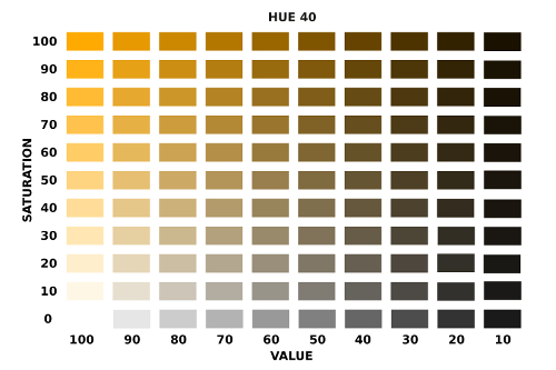
Hues are determined according to their place on a colour wheel and are numbered 0 to 360 (0 and 360 being the same hue), while saturation and value number 1 to 100 (actually decimal values between 0 and 1, but not presented as such). Thus a chart could be constructed having 3,600,000 individual colours, although the difference between individual chips would not be perceptible. The picture at right shows just such an HSV plate based on Hue 40. In this example both saturation and value have been arranged to yield a matrix of 120 chips (plus a greyscale at S=0). The plate contains more chips at this hue than many colour guides, so that mushroom collectors have a greater spread of colours in the low-saturation/high-value region. You can click on the sample plate to download a complete set.
So, should the mushroom enthusiast make up a set of HSV plates and use them to record colours? There are some drawbacks. First of all, no two computer displays will render colours identically. The colours on your laptop, tablet or cell phone may look slightly different from those on your desktop. Most modern devices are not bad, but you may wish to calibrate your monitors so they are as close in colour rendition as possible. Cell phone displays seem to be especially good with color. At low saturation (the S-component of HSV), computer monitors may not be very sensitive while cell phones work very well. For even higher accuracy you can use the Colorizer web site to fine-tune the colours between those on the plates, and there are apps that allow you to do this on your cell phone.
A second drawback to using HSV plates is that in a few years technology may change so much that programs or apps allowing the display of specific HSV colours may be extinct. Basing colour citations on the basis of published colour guides may make it easier for people reading your notes 100 years from now to see exactly what you saw. However, it is impossible to plan for the future with 100% confidence.
There are several apps for cell phones that can scan an object and report its colour directly in HSV notation. These can be useful in the field and lab but must be used with caution. The light used to illuminate the object will have a direct effect on the colour the device records, so it is necessary to adjust the "white balance" of the app to arrive at the most accurate reading. If the colour displayed on your screen differs from that of the object you may have to do some adjusting to correct it.
If all of this discussion seems a little esoteric perhaps we should get back down to what counts: preparing your specimen notes. In fact, mushroom colours vary a lot from one individual fruiting body to another. To be practical and get as many mushrooms described as possible it may suffice to write down one or two dominant colour trends and then describe briefly how this varies. Don't get carried away with too much detail or you will come home with fewer specimens. As you gain experience you will learn when you need to be exacting and when general impressions will do. And don't forget your photos!
Colour changes

Many mushrooms have tissues that change colours when cut or bruised. Sometimes this happens almost instantly after the flesh is cut, but in some species this colour change may take hours.
The mushroom at left is Russula densifolia. When it is first cut the flesh is white, but within a minute or two it turns bright red. After several hours the red disappears or darkens until the flesh is quite black. Other species of Russula related to R. densifolia may turn directly black when cut, skipping the red stage altogether. All colour changes should be carefully noted since they will not be apparent in the dried specimens and may be very important in identification.
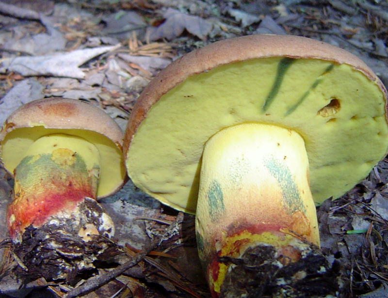

The photo above left, generously contributed by Moncton naturalist Gary Gilbert, is of Boletus speciosus one of the many boletes you may encounter. Although its colour changes are not as dramatic as those of R. densifolia they are still noticeable. The stipe is a conspicuous yellow colour with red toward the base. Both of these colours are present in the field, but the blue areas are not. These stains developed where the stipe and pore surface had been bruised during handling.
Perhaps the champion of all colour changers is Gyroporus cyanescens, pictured above right. All parts of the mushroom turn blue when cut or bruised. The flesh is white, but it is extremely difficult to get a photo of this colour because the change to blue is so rapid. The young fruiting body shown here was cut about 5 seconds before the picture was taken.
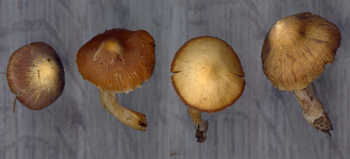
Some mushrooms are hygrophanous, that is they are very sensitive to desiccation and may change colour as their tissues dry out. This may already have happened in the field, causing you to misinterpret the colour of fresh specimens. At other times the colour fades after you collect the mushroom, as happened with the caps of a small Cortinarius shown in the picture above. The two specimens at left still retain the original colour, at least in part, while the two at right are strongly faded. Usually it is the pileus that is hygrophanous, but this may also happen to the stipe. For identification it may be important to know that the mushroom is hygrophanous and to know the colours before fading. If I find a strongly faded mushroom in the woods and am not sure about its colour when fresh I will not usually collect it.