Fleshy Fungi of New Brunswick >>
Tricholomopsis rutilans
Tricholomopsis rutilans (Schaeff.) Singer
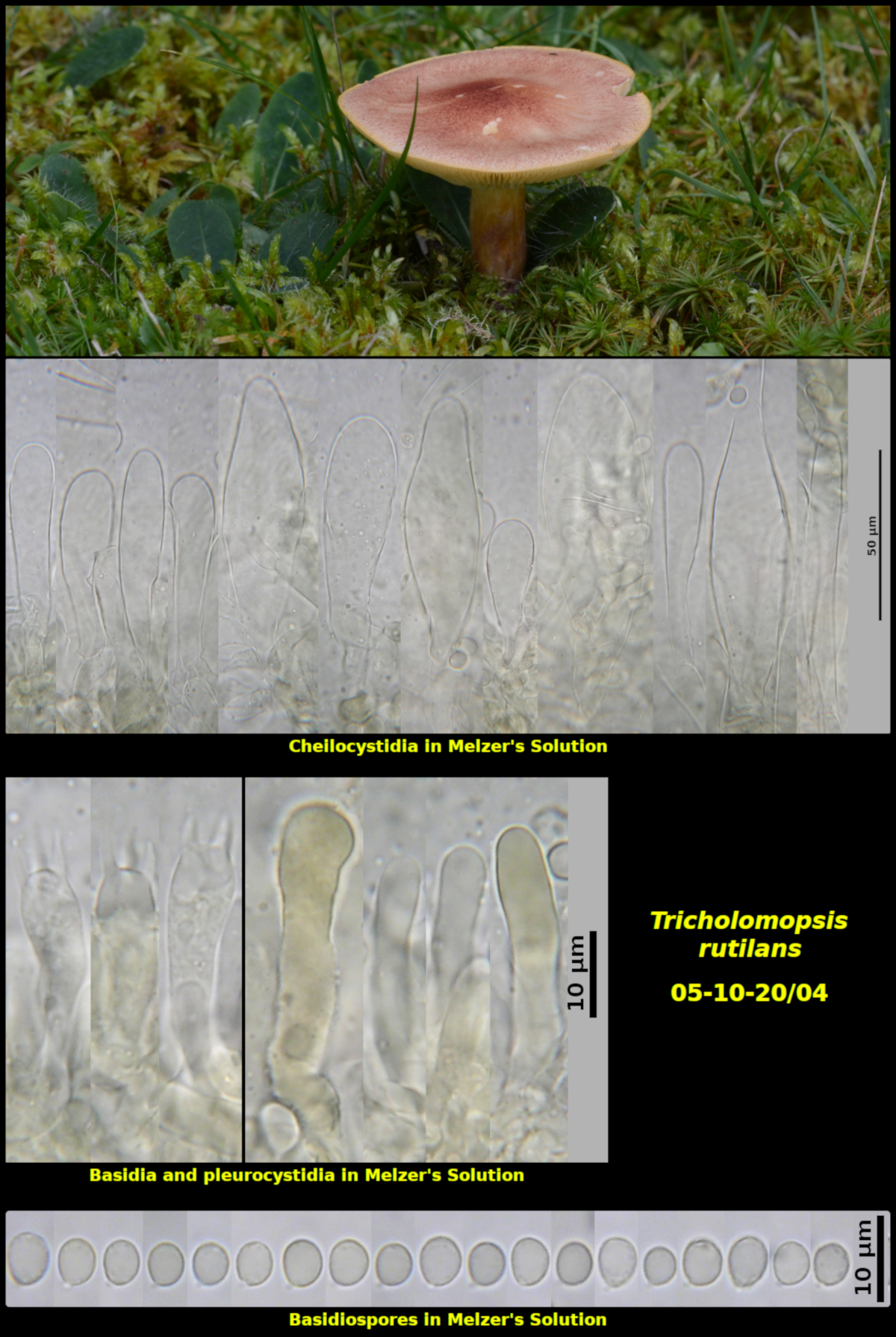
Solitary among gametophytes of Pleurozium schreiberi and Dicranum sp. in lawn, associated with Abies balsamea, Little Lepreau, New Brunswick (05-10-20/04)
Pileus not seen when young, nearly plane, with a low rounded umbo, finely appressed-scaly, with scales red to red brown (HSV350:70:60-70) over a pale yellow (HSV55:05:100) ground colour, with scales more thoroughly dominating the colour on the umbo, dry, 56 mm in diameter. Stipe equal, fundamentally yellow (HSV50:30:100) but with some fibrils in the middle having the colour of the pileal scales and influencing its colour, glabrous, dry, 53 X 10 mm. Lamellae light yellow (HSV55:05:100), close, adnate to adnexed, not marginate. Flesh pale yellow (HSV55:05:100) in the pileus and a darker lemon yellow (HSV55:50-60:100) in the stipe, lacking a distinctive odour and taste.
Basidiospores white in spore print, subglobose to broadly ellipsoidal, smooth (or very finely roughened?), unchanging in Melzer’s Solution, 4.4-6.4 x 3.8-5.3 µm, Q = 1.13-1.31 (average[41]: 5.2 x 4.3 µm, Q = 1.20). Cheilocystidia forming a continuous sterile margin, thin-walled, flaccid, variable in size and shape, cylindrical to clavate or ventricose, with a basal clamp connection, 50-102 x 10-32 µm (average[19]: 76.8 x 17.0). Pleurocystidia scattered, cylindrical to slightly clavate, with a uniform and slightly refractile content, similar to those of T. decora but less frequent, 37-40 x 5-9 µm. Pileipellis a compact cutis of colourless narrow hyphae, overlain by a surface layer of broad reddish orange hyphae. Basidia 4-spored, with a basal clamp connection, 30.4-36.4 x 7.4-8.3 µm.
Recognized by it's purple-red pileus and stipe, and by it's yellow gills and flesh. Microscopically the rather floppy and voluminous cheilocystidia and very broad inamyloid basidiospores are diagnostic.
The hymenium of Collection 05-10-20/04 was rather difficult to observe microscopically due to an abundant oily material diffusing out into the mounting medium. This material was present in lamellae mounted in water, Windex and KOH, but was immediately removed in Melzer's Solution. However, the chloral hydrate in Melzer's Solution often acts as a clearing agent, yielding very low contrast mounts difficult to observe without illumination techniques such as phase contrast or differential interference contrast (DIC).
The pleurocystidia are not especially easy to observe. They resemble those of T. decora but are not as abundant nor are they as strongly refractile.
Although often said to grow on rotting wood, a quick scan through online images of T. rutilans shows the species also to be arising from soil or forest litter. It may be that these particular basidiomata were growing on buried wood, but if so, the wood must be highly decayed. Collection 05-10-20/04 was growing in a rural lawn which had been established 17 years earlier. Another collection of this species (26-08-18/01) was found on the same lawn two years earlier but in a different place.
Photograph: D. Malloch (05-10-20/04).