Fleshy Fungi of New Brunswick >>
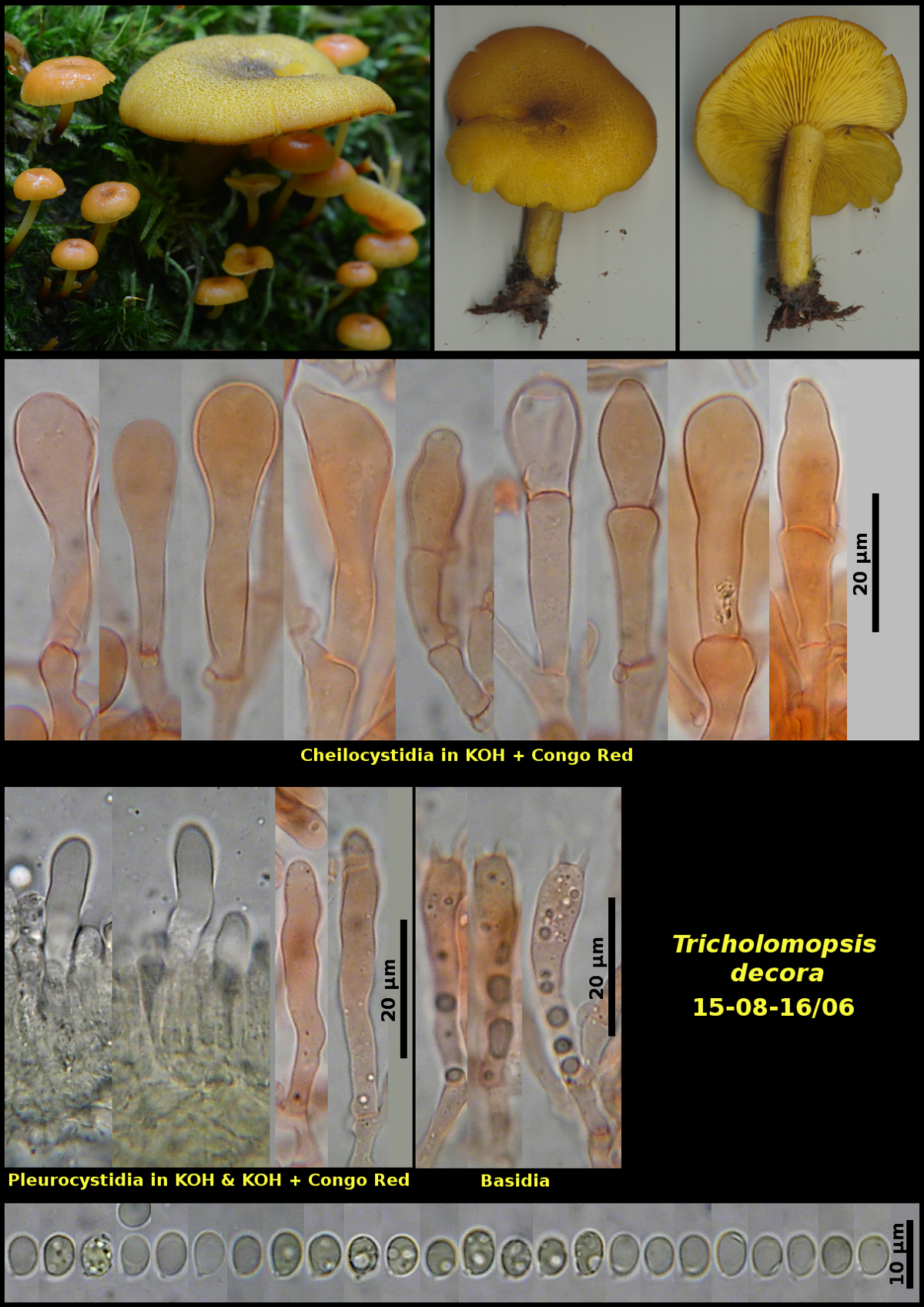
Tricholomopsis decora
Tricholomopsis decora (Fr.) Singer
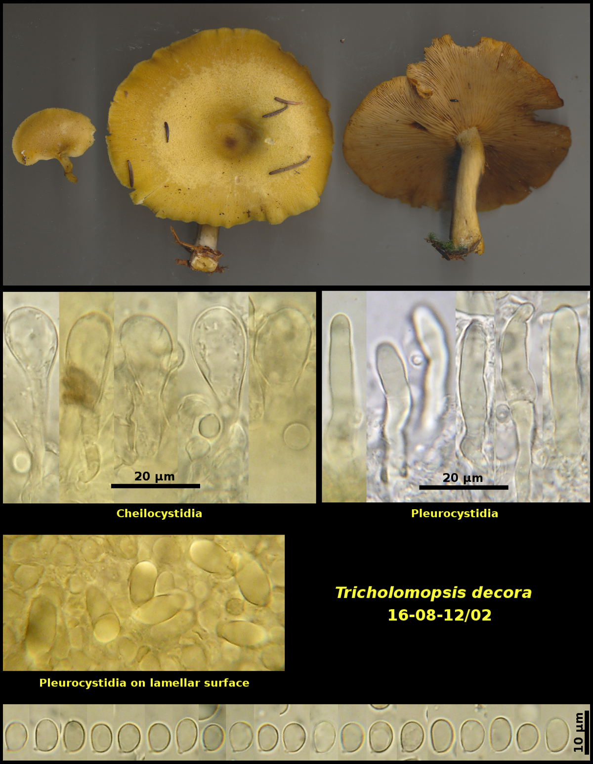
1) Gregarious on conifer log in forest of Picea rubens, Abies balsamea and Betula papyrifera, Caledonia Gorge Protected Natural Area, New Brunswick (16-08-12/02)
2) Solitary amongst basidiomata of Xeromphalina campanella on a dead conifer log in mixed forest of Acer saccharum, Betula alleghaniensis, B. papyrifera and Abies balsamea, Nepisiguit Protected Natural Area, New Brunswick (15-08-16/06).
Basidiospores ellipsoidal, hyaline, smooth, without iodine reactions, 6.1-7.8 X 4.6-5.5 μm, D/d 1.23-1.56 (average[23]: 6.9 X 5.1 μm, D/d = 1.36)(16-08-12/02)
Basidiospores white in spore print, broadly ellipsoidal to obovoid, hyaline, smooth, without iodine reactions, 5.1-6.6 X 4,1-4,9 µm, D/d = 1.15-1.50 (Average[27]: 5.9 X 4.4 µm, D/d = 1.33)(15-08-16/06)
Although some authors have called attention to the refractile cells in the hymenium of T. decora and called them pleurocystidia, others state that all members of the genus lack them. These cells are particularly abundant in 16-08-12/02 and are here labeled as pleurocystidia.
The two collections described and figured here appear to fit current concepts of T. decora. However, there are some differences both macro- and microscopically. Collection 16-08-12/02 has fewer and less conspicuous scales on the pileus, stronly clavate cheilocystidia and larger basidiospores. Collection 15-08-16/06 has conspicuous dark scales on the pileus, prominent but rather variable cheilocystidia and smaller basidiospores. These two collections, along with some others not yet studied in detail would benefit from genetic sequencing.
Photograph: D. Malloch (16-08-12/02, 15-08-16/06).