Essays >> Cleistothecial Ascomycota >> Cleistothecial Ascomycota - Plate 04
Cleistothecial Ascomycota - Plate 04

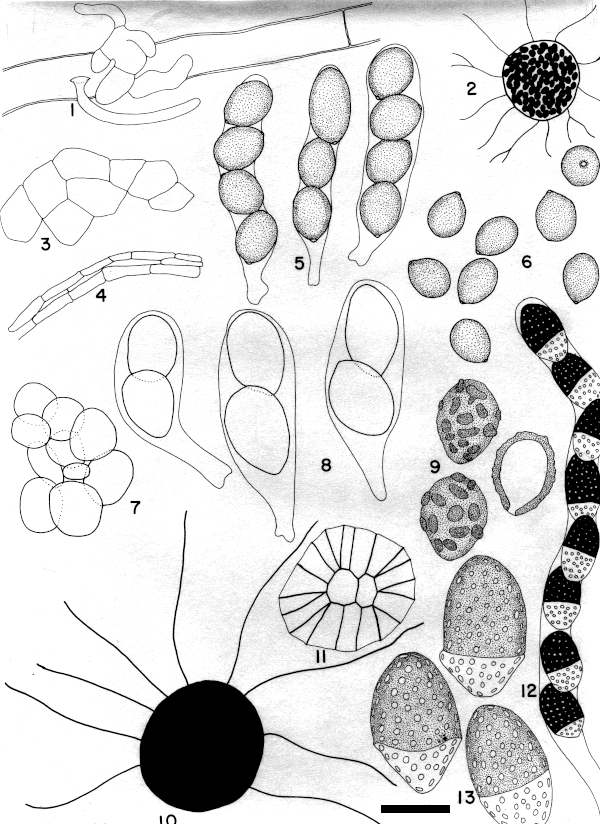
1-6. Boothiella tetraspora Lodhi & Mirza
1. Ascomatal initials (X 1900). 2. Ascoma (X 165). 3. Surface of peridium (X 825). 4. Cross section of peridium (X 825). 5. Asci (X 1900). 6. Ascospores (X 1900).
7-9. Copromyces bisporus N. Lundq.
7. Surface of the peridium (X 1900). 8. Asci (X 1900). 9. Ascospores, including one in optical section (X 1900).
10-13. Diplogelasinospora princeps Cain
10. Ascoma (X 165). 11. Cephalothecoid peridial plate (X 1900). 12. Ascus (X 825). 13. Ascospores (X 1900).
At least for now, all the species on this plate still remain in their original genus and all are still considered to be members of the Lasiosphaeriaceae or Sordariaceae.
Boothiella tetraspora has hyaline ascomata that appear dark when the ascospores mature. The ascospres are relatively thin-walled, are easily broken and have a single large germ pore. In culture on V8 agar, the colonies grow rapidly, filling the plate in three days at 30 C and producing mature ascomata in less than 2 weeks. It is known from dung and soil. The illustrations here are from TRTC 38871, from donkey dung in Pakistan, and CBS 151.68, a culture derived from the holotype of Thielaviella humicola, a later synonym of Boothiella tetraspora.
Copromyces bisporus is immediately recognizable by its two-spored asci and brown warted ascospores with a single germ pore. It is known from both Europe and North America on dung of rabbits, rats and cows. Petrak (Sydowia 21: 241. 1968) believed it to belong in the genus Rechingeriella and transferred it there as R. bispora. However, Rechingeriella is gconsidered to be a member of the unrelated family Zopfiaceae and Petrak's transfer is not generally accepted. The illustration here of C. bisporus is from TRTC 36559, a collection of cow dung from San Luis Potosí, Mexico.
Diplogelasinospora princeps has ascospores pitted like those of many Neurospora species, but differs in having two-celled ascospores, with one cell dark and one hyaline. Similar to Neurospora species, D. princeps grows rapidly in culture but differs in producing ascomata rather slowly. There are presently four species of Diplogelasinospora known. García, Marín and Cano (Persoonia 32: 286-287. 2014), describing one of these, D. moalensis, provided a cladogram based on ITS sequences for all four, and briefly discussed their relationships. Cai, Jeewon and Hyde (Mycol. Res. 110: 137–150. 2006.), in an extensive phylogenetic investigation of the Sordariaceae, concluded that Diplogelasinospora is closer to the Lasiosphaeriaceae than the Sordariaceae.
Diplogelasinospora princeps was orginally described from flax seed in Canada, while the other three are known only from soil. The illustration here is based on cultures derived from TRTC 37076, the holotype.