Fleshy Fungi of New Brunswick >>
Inocybe aurea
Inocybe aurea Huijsman


Four collections, three illustrated here:
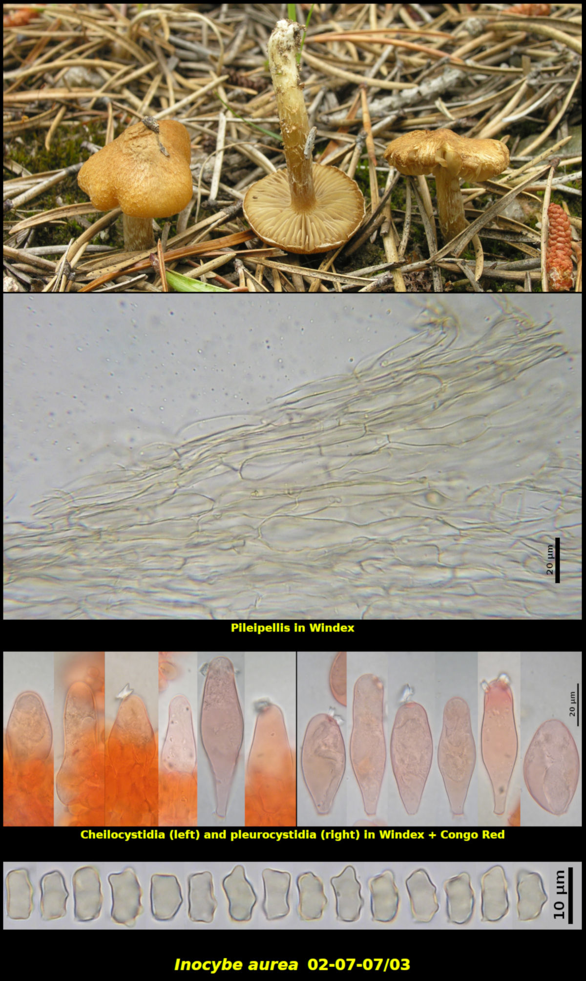
1. Gregarious to clustered in needle litter of Pinus banksiana in sandy disturbed soil. The pines were fairly large and part of a plantation, N of Lepreau, Charlotte Co., New Brunswick (02-07-07/03). - The entire plantation was completely harvested a few years later.
Pileus conic-convex at first, expanding to convex or plane at maturity, with a small and often inconspicuous umbo, dry, becoming somewhat cracked at centre and minutely squamulose-scaly, elsewhere becoming more appressed-fibrillose to rimose, Pale yellow to Light yellow (Methuen 3-4A3-4) when young, later developing more orange-brown shades near Grayish orange (5B4), 12-27 mm in diameter. Stipe white at first, later developing some dull shades near Orange gray (5B2), equal, dry, finely fibrillose with some veil fragments, equal or tapering down slightly, 16-37 X 2-5 mm. Lamellae white at first, darkening as the spores mature, close, adnexed, not marginate. Cortina white. Flesh white in both pileus and stipe, lacking a distinctive odour.
Basidiospores reddish brown in spore print, nodulose, with 3-5 nodules in profile and 6-8 in dorsi-ventral view, with nodules difficult to count but probably 8-12, characterized by apical nodules usually nearly equal in height and rather low lateral nodules giving an overall rectangular appearance in profile, 8.1-11.0 x 4.6-6.4 μm, Q = 1.41-2.03 (average[27]: 9.2 x 5.5 μm, Q = 1.69). Pileipellis a layer several cells deep of repent parallel lightly encrusted clamped reddish brown hyphae 4-19 μm in diameter. Cheilocystidia forming a continuous to somewhat discontinuous sterile margin, ventricose to sublageniform, inequilaterally obovoid when young, thin-walled, hyaline in 5% KOH, often with large apical crystals, clamped at base, 39-71 X 15-25 μm, accompanied by less structurally uniform clavate frequently orange brown paracystidial elements measuring 29-74 X 9-23 μm, with paracystidia forming a nearly continuous sterile margin. Pleurocystidia abundant, lageniform to clavate, thin-walled, usually abruptly truncate at base and without a stipe, clamped at base, hyaline and remaining so in 5% KOH, (36-)54-76 X 14-22(-31) μm. Caulocystidia produced only at the apex of the stipe, variable in size and shape, varying from clavate to broadly ventricose to nearly cylindrical, rarely slightly lageniform, usually with thin walls but sometimes with walls uniformly thickened to 0.8 μm, 20-87 X 7-18 μm, replaced by the hyphal vestiture below the apex. Basidia clavate, 4-spored, clamped at base, 26-34 X 8.4-10.0 μm.
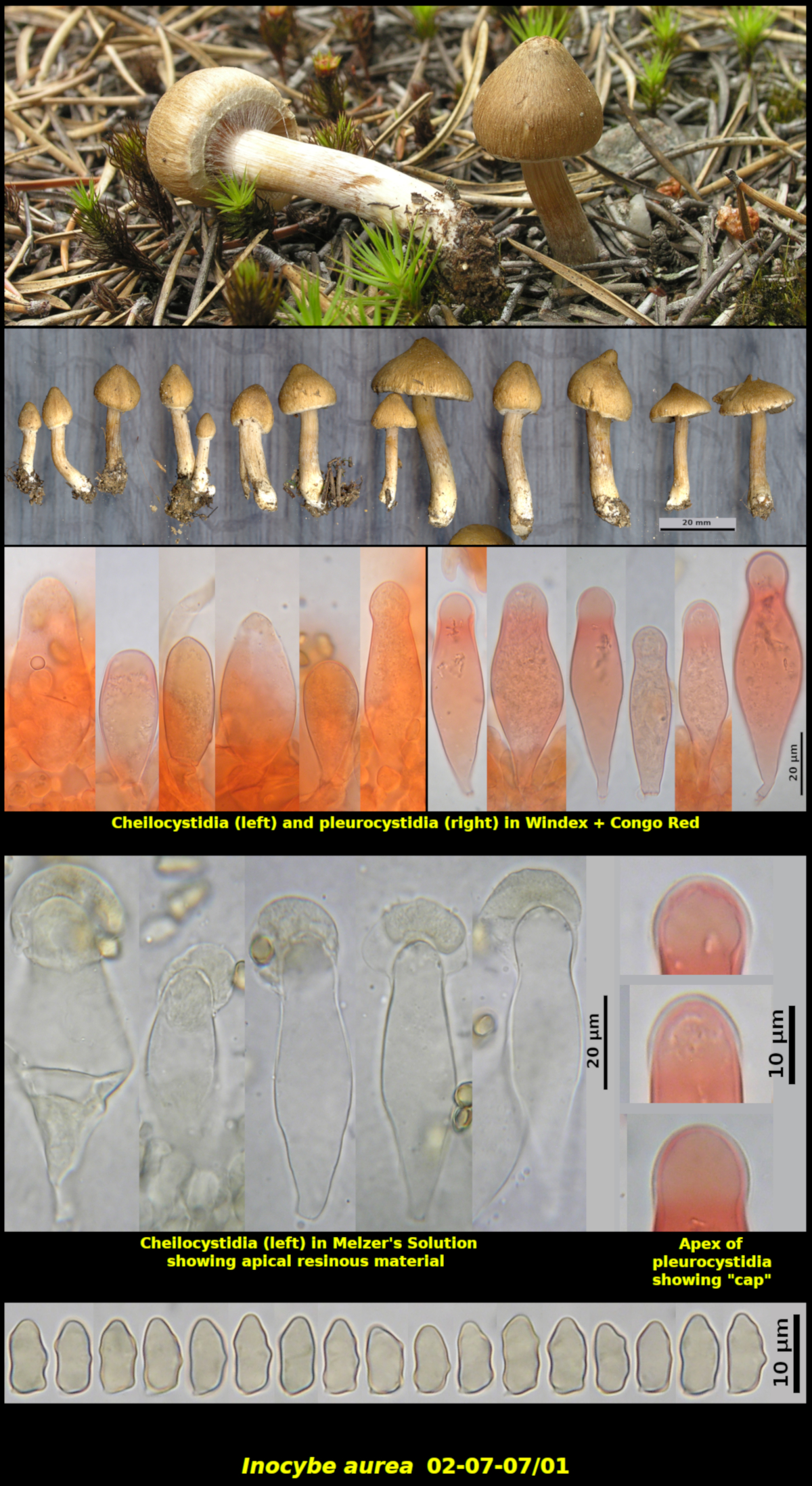
2. Gregarious to clustered in needle litter of Pinus banksiana in sandy disturbed soil. Same locality as above. (02-07-07/01).
Pileus conic-turbinate at first, expanding to more broadly conical and then undulate-plane, with a broad to fairly sharp and prominent umbo, dry, glabrous to radially appressed-silky-fibrillose, with fibrils becoming more pronounced and rimulose in older specimens, with some white veil fragments at the margin, Grayish orange to Brownish orange (5BC3), darker on the disc, darkening in older drying individuals (especially on the umbo), 12-33 mm in diameter. Stipe Grayish orange (5-6B3), nearly white at the base, equal or slightly swollen below, dry, solid, almost peronate with appressed white veil material at first and with remaining at base, appearing glabrous but with some fine white-fibrillose streaks of veil fragments elsewhere, 32-53 X 3-6 mm. Lamellae nearly white to Orange gray (5B2) at first, darkening as the spores mature, close, adnexed, not marginate. Cortina white. Flesh white in the pileus and more or less concolorous with the surface tissues in the stipe, lacking a distinctive odour although massed basidiomata have a faint odour resembling shrimp or some sort of seafood or perhaps more like Lycoperdon spp., tasteless.
Basidiospores orange brown in spore print, nodulose, with >10 low nodules, often more wavy in outline than nodulose, 8.6-11.2 X 4.4-5.3 μm, Q = 1.70-2.27 (average[39]: 9.6 x 4.8 μm, Q = 1.99). Pileipellis a layer several cells deep of repent parallel lightly encrusted clamped reddish brown hyphae 4-19 μm in diameter. Cheilocystidia abundant, mostly ventricose, less commonly lageniform, thin-walled, with walls up to 1.6 μm thick, without apical crystals, with an apical cap up to 1.1 μm thick that appears to absorb less Congo Red stain than the walls, occasionally with a thick resinous dome up to 9 μm deep when mounted in Melzer’s Solution, 41-70 x 16.3-29 μm. Pleurocystidia abundant but scattered, mostly lageniform but often sublecythiform, thin-walled, with walls (rarely) up to 1.7 μm thick, with an apical cap up to 1.5 μm thick that appears to absorb less Congo Red stain than the walls, 53-78 x 14-24 μm.
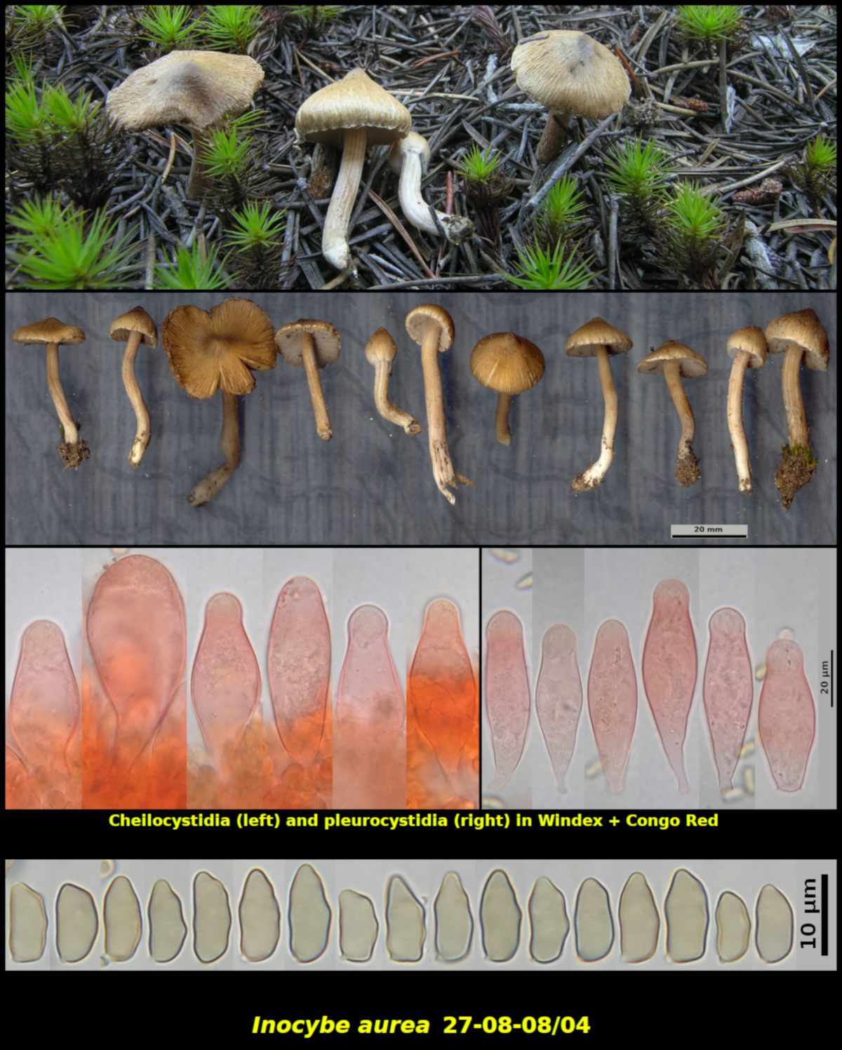
3. Gregarious (many) in sandy soil under Pinus banksiana. Same locality as above but one year later and later in the season (27-08-08/04).
Pileus conical at first, expanding to more broadly so at maturity, with a fairly sharp umbo, radially silky-fibrillose, not becoming scaly, faintly rimose at the margin at first but becoming more markedly so in age, Greyish brown (7EF3), Brownish grey (5C3) toward the margin, with some whitish veil fragments at the margin, 12-32 mm in diameter. Stipe Greyish orange to Orange grey (5B2-3), equal, dry, glabrous and apparently not pruinose, 22-42 X 3-4 mm. Lamellae white, close, adnexed, not marginate. Cortina white, attached near the apex of the stipe. Flesh white in the pileus and faintly yellowish white in the stipe, with a faint mushroom odour.
Basidiospores not forming a visible spore print, nodulose with more than 10 nodules, with nodules low, rounded and forming a wavy outline, with lower nodules more prominent in dorsi-ventral view, 8.5-12.9 x 4.4-6.7 μm, Q = 1.72-2.52 (average[34]: 10.6 x 5.1 μm, Q = 2.09). Cheilocystidia abundant, ventricose to lageniform, thin-walled, with walls up to 1.0 µm thick, without apical crystals, hyaline in KOH, 56-84 x 15-37 μm. Pleurocystidia abundant, scattered, similar to the cheilocystidia but more consistently lageniform, thin-walled, with walls up to 1.1 μm thick, without apical crystals, hyaline in KOH, 54-76 x 15-26 μm.
Inocybe aurea appears to be a rare species, reported up to the now mainly from Europe. Originally described from The Netherlands, it has been reported from Germany and Switzerland. It is included in Funga Nordica where it is stated to be rare. The website for the Herbarium of The Natural History Museums and Botanical Garden, University of Oslo (http://www.nhm.uio.no/botanisk/sopp/redgroup.htm) lists I. aurea as rare in its Redlist of Threatened Fungi of Norway.
Horak (Arctic and Alpine Mycology II, 1987) provided a short description of I. aurea in his key and a plate of excellent line drawings, including four spores from Huijsman’s type. The microscopic features of our New Brunswick material are mostly in accordance with Horak’s plate and description.
One point of disagreement between our material and Horak’s concern the plant associations. The three Swiss collections were from snow beds at an elevation of 2350-2420 m and grew in association with Salix herbacea, while the New Brunswick material was from a sandy pine plantation. Huijsman’s type was collected in dry sandy pine woods in The Netherlands, a habitat and elevation much closer to that of the New Brunswick collection. The greatly different habitat and plant association of the Swiss material suggests that it could be a species other than I. aurea.
The New Brunswick collections of I. aurea are characterized by 1) the yellow to yellow brown fibrillose pileus, 2) thin-walled and broad cheilo- and pleurocystidia and 3) narrow nodulose basidiospores. Inocybe ventricosa Atkinson is probably very similar. Grund and Stuntz (Mycologia 73: 655-674. 1981), who examined two collections of that species from Nova Scotia as well as the type from New York, described the pileus as yellow ochre and having a broad rounded umbo and a silky to rimose surface. The cheilo- and pleurocystidia are ventricose, thin-walled and similar in size to those of I. aurea. The only significant difference between I. ventricosa and I. aurea appears to be in the basidiospores. Those of I. ventricosa are described and illustrated by Grund and Stuntz (1981) as (6.5-)7-8(-9) X (4.5)5-6(-6.5) μm and thus shorter but proportionately broader than those of I. aurea. Unfortunately Grund and Stuntz did not provide very detailed information about plant associates (“Solitary on soil in moss, in hardwood forest or mixed stand…”) but their collections may not have come from sandy pine woods as have the New Brunswick and type collections of I. aurea The herbarium at Acadia University (ACAD) has uploaded to MycoPortal a record of I. aurea, collected by H. Stewart near Aldersville, Nova Scotia in 1966 and identified by Dr. Grund. Oddly, the series of papers on Nova Scotian Inocybe species by Grund and Stuntz make no reference to this collection.
Although collected in the same locality in sandy soil under Pinus banksiana, the four collections from New Brunswick differ from each other in some respects, most notably in the colour of the pileus, presence or absence of apical crystals on the cystidia and in the appearance of the basidiospores. Collection 02-07-07/03 is closest to the type as presented by other authors. The other New Brunswick collections have a darker and less overtly yellow pileus. The basidiospores of 02.07-07/03 are more distinctly nodulose than the others. However, these features may only represent the natural variation among basidiomata from the same location, perhaps affected by age and season.
Photo: D. Malloch (19-08-16]01).