Home >> Diversity and classification >> True fungi >> Dikarya >> Ascomycota >> Pyrenomycetes >> Laboulbeniomycetes
THE LABOULBENIOMYCETES
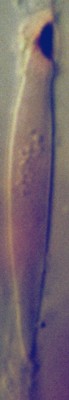
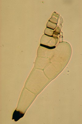
The Laboulbeniomycetes form a distinct and unusual group of fungi. All are parasitic on insects and related arthropods but remain on the outside of the animal in much the same way a tick or leech would. The feature all Laboulbeniomycetes have in common is the ascospore. The photo at left shows a typical ascospore of the Laboulbeniomycetes. The spore is long and narrow and has a dark attachment on the upper end. Although the photo does not show it there is a septum near the middle of the spore. The ascospores are not forcibly discharged from the ascus but instead collect inside the perithecium and then are pushed up out of the ostiole. They collect at the ostiole in a cluster or in a chain, attachment pad up, and adhere to a mite or insect when it comes in contact with them. They adhere to the animal by the attachment pad and then penetrate its cuticle with an absorption cell called a haustorium. Once the haustorium is in place the spore can begin to develop into its final form.
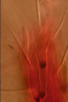
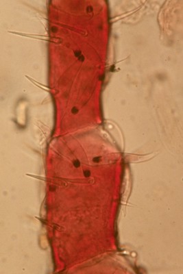
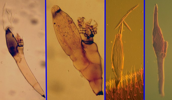
The two pictures at right show a species of Pyxidiophora in the early stages of colonizing a mite. The large pink structure running the length of the leftmost picture is the front leg of the mite. The dark spots along the leg are the attachment pads on the ascospores of the Pyxidiophora species. Looking closely you can see the rest of the spore extending away from the pad. The picture at far right is of the end of the first leg of another mite with four ascospores attached. The most important part of this picture is the ascospore furthest to the left which is producing a single spore at its tip. This is important because unlike other fungi that may be transported by insects and mites, members of the Laboulbeniomycetes tranform their ascospores into spore-bearing structures while attached to the animal and, because of the nutrition obtained through the haustorium, are able to produce significant numbers of spores. The extent to which this attached spore develops forms the basis of the two orders, the Pyxidiophorales and the Laboulbeniales.
THE PYXIDIOPHORALES
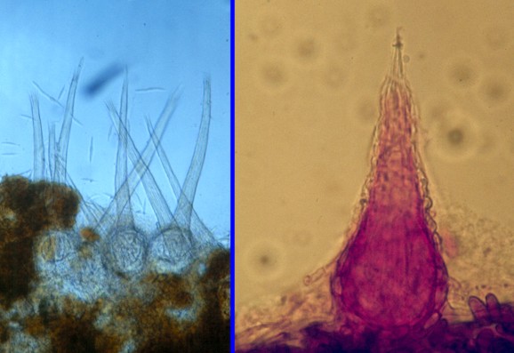

In many ways the Pyxidiophorales are fairly conventional pyrenomycetes, producing perithecia and ascospores on a variety of substrates. The picture at far left shows perithecia of two species, the one in the left panel on dung, the one at right on rotting seaweed. The colours are not true-to-life; species of Pyxidiophora have pale, almost colourless perithecia but these photos were taken of material prepared for microscopic examination using blue and pink stains. The two species obviously differ in the length of the perithecial necks, one very long and one very short. Since the ascospores are exposed for dispersal at the perithecial ostiole the length of the neck is probably critical; long-necked species could inoculate taller mites or insects than shorter ones. However, the ascospores sometimes collect in long chains extending up from the ostiole, as in the other picture at near-left, so it may be that neck length is not the whole story. Neck length varies from species to species in all habitats. Thus the short-necked one is not particularly characteristic of seaweed, long-necked ones can be found there too.
The perithecia of Pyxidiophora are usually quite small, less than 1 mm tall including the neck, colourless and often buried partly in the substrate. Although they are very common in materials undergoing decomposition they can be difficult to find. They usually appear early in the decomposition process and remain for less than a week. You must be there at the right time to find them and use a good binocular dissecting microscope to do so.
Ascospores of the Pyxidiophoraceae probably do not remain long on their animal host.
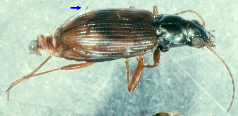
THE LABOULBENIALES As far as we know, the Laboulbeniales is much the larger of the two orders of Laboulbeniomycetes. The great ecological success and diversity of this order probably stems from the fact that no other group of organisms has successfully occupied their habitat. They differ from the Pyxidiophoraceae in one particular attribute; their life history has been compressed so drastically that they live their entire lives on the surface of insects and mites without a more conventional mycelial stage. The compression is so great that even their perithecia are produced on the animal. The picture at left shows an ascospore of Laboulbenia philonthi that has already developed a complexity beyond anything seen in the Pyxidiophoraceae. The attachment pad at the base of the modified ascospore is still present but the rest of it has undergone several cell divisions and modifications. The sack-like structure on the right side of the spore is the beginning of a perithecium. The left part of the ascospore shows some development into what will become appendages extending out beside the perithecium. Many species may start producing spermatia, spore-like cells with a fertilization role, at this stage. The mature stage of ascospore development, including the perithecium, spermatial, appendages is called the "thallus" (plural: thalli) by most authors. The picture at right illustrates four species of Laboulbeniales at maturity. The panel furthest to the left portrays a mature thallus of Laboulbenia philonthi, the species illustrated in its immature stage above left. The large dark structure is the perithecium, which has already begun to discharge its ascospores. The appendages are now below the apex of the perithecium. The two panels to the right of L. philonthi are also of species of Laboulbenia, L. cafii, and an unidentified species. All three species were collected on the bodies of members of the beetle family Staphylinidae within piles of rotting seaweed. The thallus at far right is from a species of Stigmatomyces, most commonly found on flies. So far you may not have a very good idea of how large the thalli of the Laboulbeiales might be. In fact they vary quite a bit in size but are mostly quite small. The picture above shows a species of Bembidion, a ground beetle, bearing a single thallus of Laboulbenia vulgaris, indicated by a blue arrow. The entire beetle is only about 5 mm in length, so the fungus is indeed very small. Often an insect will have only a single thallus attached but at times they may become heavily infested. It is not known what effect the fungus has on the insect, but we can imagine that a large infestation might affect the flight of the insect or drain it of nutrients. It is thought that species of Laboulbeniales may be spread from insect to insect during mating. This idea is supported by the fact that species of Laboulbeniales have a very narrow host range and often occur on a very particular site on the insect. Being structurally simple fungi it is not surprising that they are most common on insects living in moist environments. They are most easily found on insects living in composting plant materials or in wet or even aquatic habitats. Water beetles can be quite productive. Although they occur on a great variety of insects, beetles, particularly ground and rove (Carabidae and Staphylinidae), are especially good sources.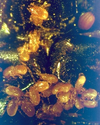
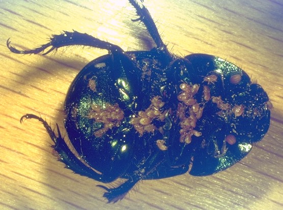 Those that have been studied were found on mites that hitch a ride on dung beetles as a means of travelling from dung pile to dung pile. The pictures at right illustrate this: a large dung beetle has been turned on its back to reveal a crowd of yellowish-brown mites clinging to its underparts. The one furthest right shows a close-up of the mites, holding on by their chelicerae (pinchers) with their legs dangling in the air. Some of these mites undoubtedly carry ascospores of Pyxidiophora species as well as spores of other fungi. The beetle itself may also serve as a more direct means of transport for some fungi. Drs. Meredith Blackwell and David Malloch, working in Algonquin Provincial Park in Ontario, described several patterns of ascospore development on mites. Some ascospores remained fairly simple and produced single spores at their tips, as in the photo above, while others produced rather elaborately modified structures bearing several spores. When the beetle arrives at a new pile of dung the mites drop off and begin moving about in search of food, and as they do, deposit the newly-formed spores of Pyxidiophora.
Home >>
Diversity and classification >>
True fungi >>
Dikarya >>
Ascomycota >>
Pyrenomycetes >>
Laboulbeniomycetes