Fleshy Fungi of New Brunswick >>
Tricholoma virgatum
Tricholoma virgatum (Fr.) P. Kumm.
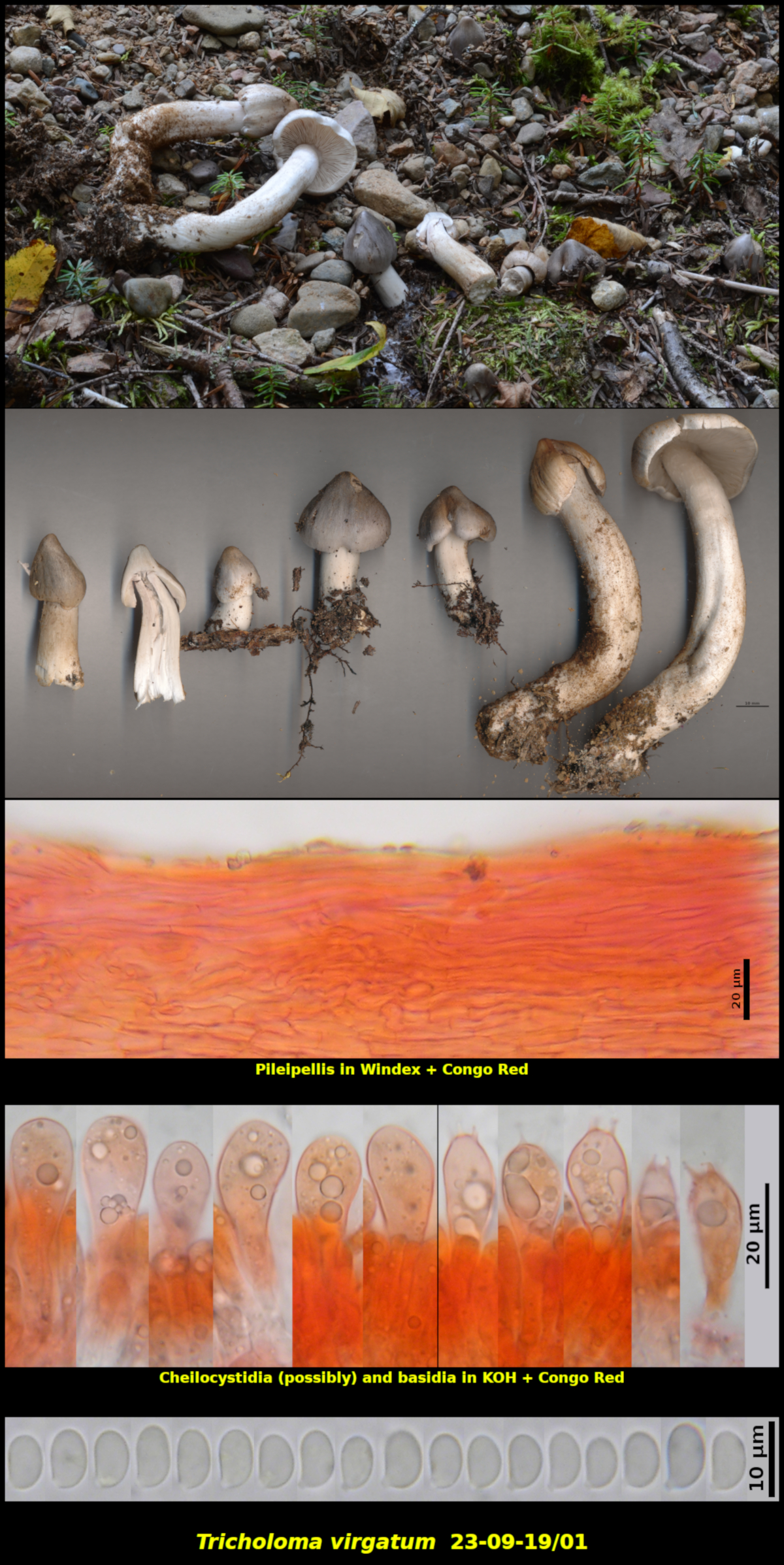
Gregarious (6-10) in gravel and forest soil at the edge of a gravel driveway, associated with Abies balsamea and Betula cordifolia, Little Lepreau, New Brunswick (23-09-19/01).
Basidiospores white in spore print, ellipsoidal to phasaeoliform, usually with a suprahilar depression, smooth, without colour changes in Melzer’s Solution, 6.5-8.5 x 4.1-5.0 μm, Q = 1.46-1.83 (average[36]: 7.3 x 4.5 μm, Q = 1.62). Basidia 4-spored, without a basal clamp connection. Cheilocystidia possibly present as scattered clavate cells but these may only be immature basidia. Pileipellis a cutis of narrow hyphae over a subcutis of slightly broader hyphae, without a pseudoparenchymatous subcutis, with upper cuticular hyphae perhaps subgelatinous but this is not obvious. Clamp connections lacking on all hyphae of the basidiomata.
Tricholoma virgatum is characterized by its silvery grey sharply umbonate pileus and white bitter to acrid flesh. As its name "virgatum" implies, the pileus is radially streaked or virgate with appressed fibrils, although this characteristic may not be strongly expressed. Microscopically it is characterized by a lack of both clamp connections and gelatinous tissues.
Collection 23-09-19/01 was growing beside a rural driveway that had recently been resurfaced with gravel. The new gravel must have partially covered an established colony of the fungus, forcing it to produce long stipes to reach the surface. Basidiomata produced in the area not covered by gravel had short stipes.
The basidiospores of Collection 23-09-19/01 are markedly narrower than those given for basidiospores of T. virgatum in both the European and North American literature, although the lengths are comparable. It is impossible to draw any taxonomic conclusions based on this single collection but it might be interesting to examine this discrepancy in other collections of the species. There is some mention in the literature of cheilocystidia in T. virgatum, probably in reference to the structures shown above. However, these closely resemble developing basidia, leaving some doubt about their true nature. Note also in the picture above the last two basidia on the right which are smaller than the others and probably remained post-discharge.
Photograph: D. Malloch (23-09-19/01).