Fleshy Fungi of New Brunswick >>
Mycena radicatella
Mycena radicatella (Peck) Sacc.
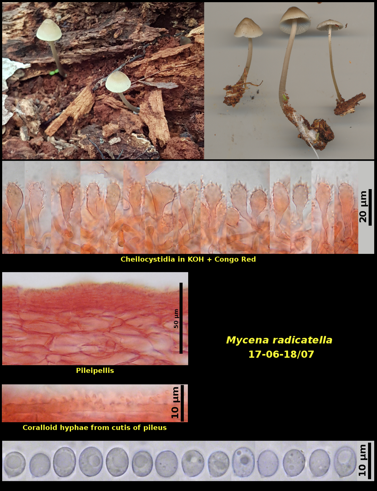
Gregarious (2) on decayed hardwood log in beech-maple forest, Spednic Lake Protected Natural Area, York Co., New Brunswick (17-06-18/07).
Basidiospores white in spore print, broadly ellipsoidal to obovoid to nearly globose, smooth, strongly amyloid in Melzer’s Solution, 6.5-8.3 X 5.6-6.6 µm, D/d = 1.09-1.34 (average[27]: 7.4 X 6.0 µm, D/d = 1.22). Cheilocystidia forming a sterile margin, clavate, covered with finger-like projections. Pleurocystidia lacking. Pileipellis a rather dense cutis of narrow hyphae overlying the broader hyphae of the pileus trama, with coralloid hyphae on the surface
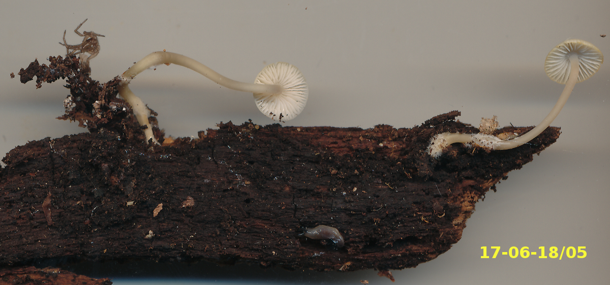
Recognized by its pale pileus and growth on wood of hardwoods. Although Collection 17-06-18/07 does not show it especialy well, the stipe is often long and rooting down into the wood. Collection 17-06-18/05, found on a differnt log at the same locality, is more typical for this feature. Microscopically the species is distinguished by its cheilocystidia with finger-like projections and its broad basidiospores. Mycena cf. galericulata, found in the same area and on the same day, differs in its growth on the ground rather than on wood and in the structure of the upper part of its pileus. A cross section of the pileipellis of that species shows the tissues immediately below the coralloid pellis as composed of broad hyphae seated above much narrower hyphae. In Collection 17-06-18/07 the subpellis is composed of narrow hyphae seated above much broader hyphae. Mycena atroalboides is similar both macro- and microscopically but occurs on the ground among mosses and has more ellipsoidal spores.
Photograph: D. Malloch (17-06-18/07).