Fleshy Fungi of New Brunswick >>
Mycena purpureofusca
Mycena purpureofusca (Peck) Sacc.
Two collections:
-
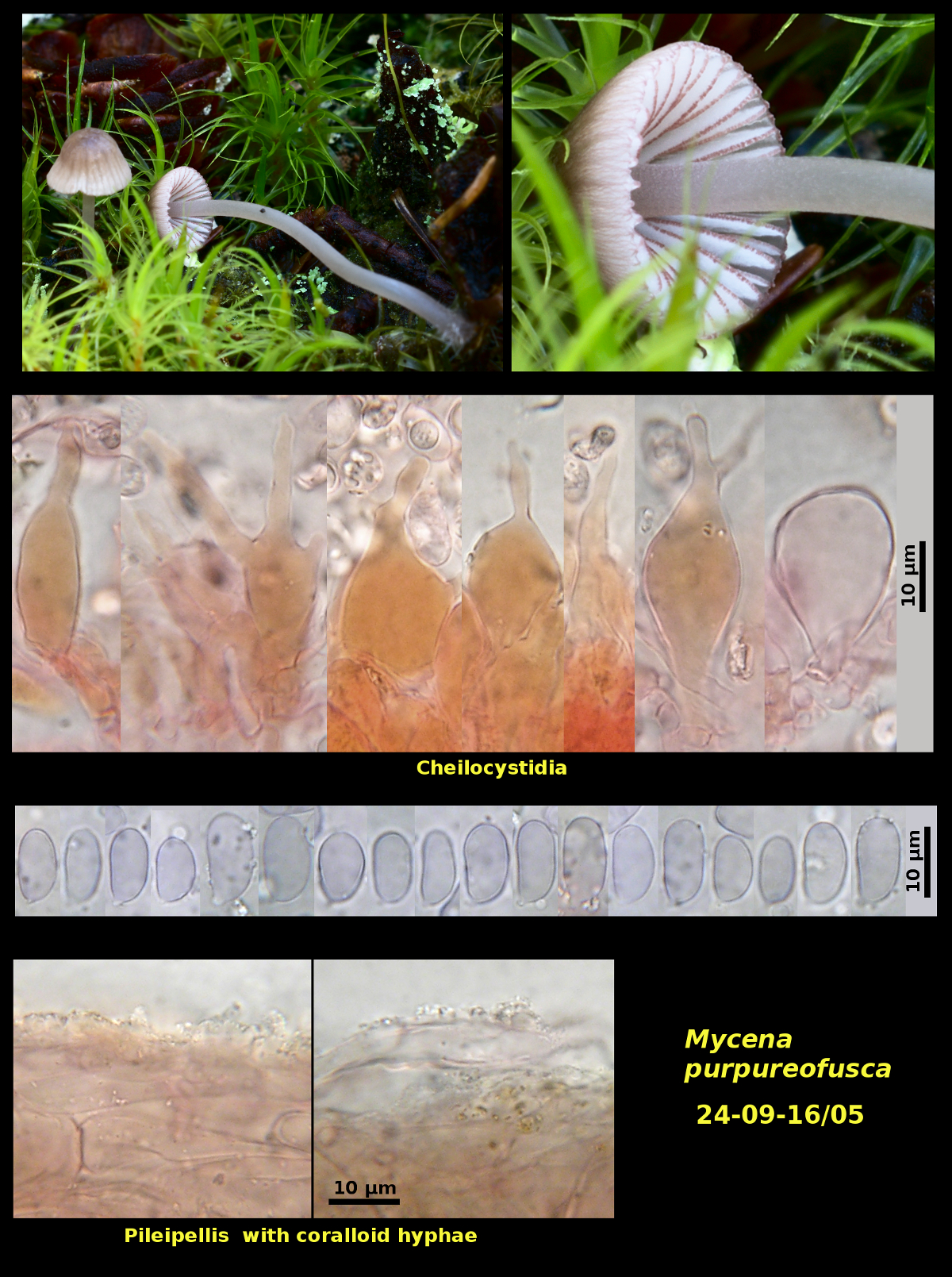
Scattered on fallen twigs of Abies balsamea in forest dominated by Abies balsamea and Betula cordifolia, Campobello Island, New Brunswick (24-09-16/05).
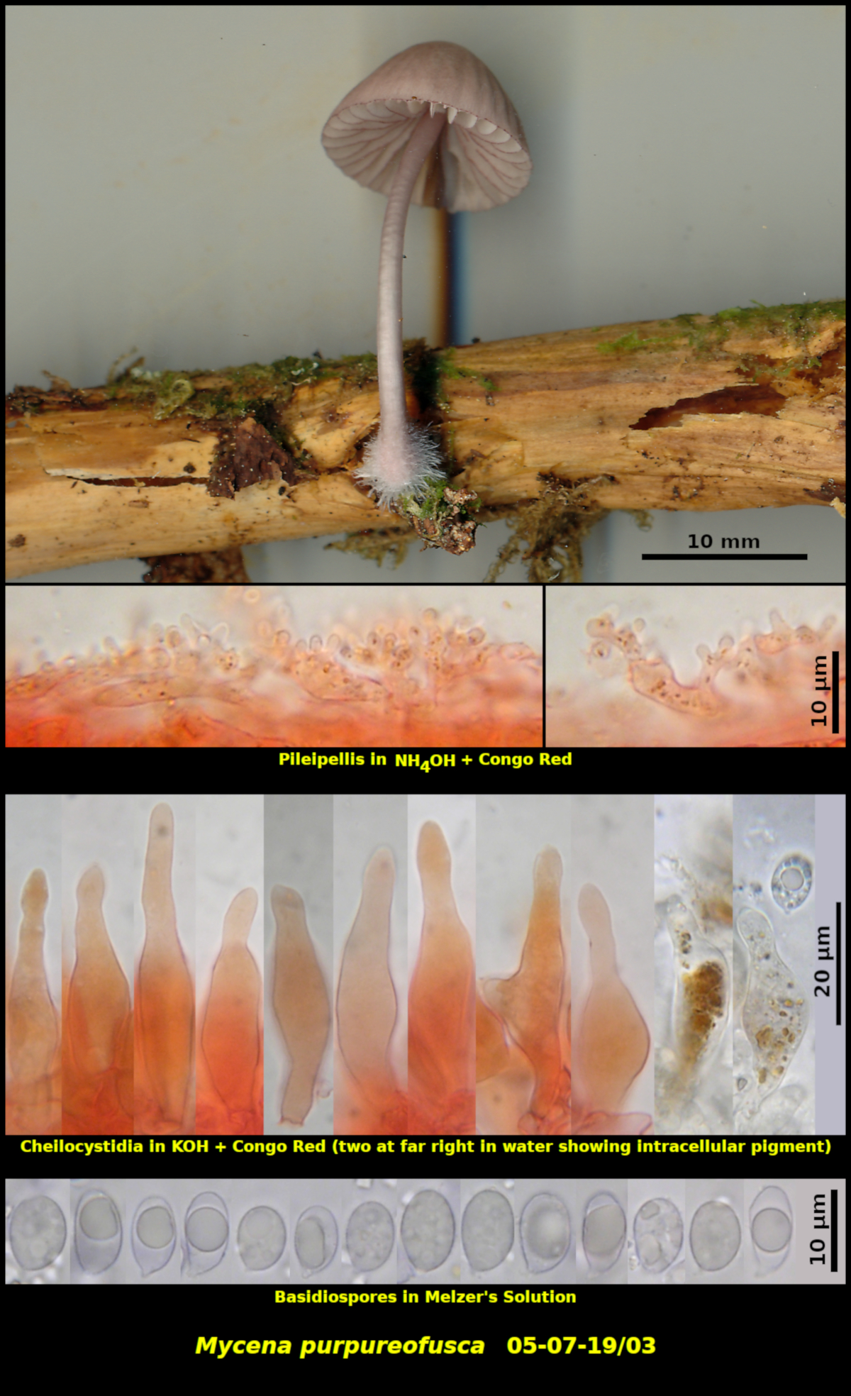
Solitary on a fallen and decaying conifer branch in a wet area in forest dominated by Picea rubens, Abies balsamea and Betula papyrifera. Upper Dungarvon Protected Natural Area, New Brunswick (05-07-19/03)
Basidiospores white in spore print, ellipsoidal to narrowly obovoid, smooth, amyloid, 7.9-12.4 x 4.5-6.9 μm, Q = 1.29-2.29 (average[42/2]: 9.9 x 5.7 μm, Q = 1.75). Cheilocystidia forming a continuous sterile margin, irrgeularly ventricose to lageniform, occasionally to frequently lobed or branched, containing a brown granular pigment in dried material, 27-53 X 7.8-17.0 µm. Pleurocystidia scattered, most abundant toward the edge of the lamella, similar to the cheilocystidia. Basidia 4-spored, clamped at the base. Pileipellis a thin cutis of coralloid hyphae above a subpellis of broad hyphae. Clamp connections present but often difficult to observe.
Collection 24-09-16/05 has rather more branched cheilocystidia than those of 05-07-19/03. According to Smith’s illustrations (North American Species of Mycena, 1947) this would place it in M. purpureofusca and 05-07-19/03 in M. elegantula sensu Smith. The difference in fruiting time (early July vs. late September) might support the separation.
Perry & Desjardin (Mycotaxon 70: 87-97. 1999) examined the types of M. elegantula and M. californiensis, both holotypified from California collections, and concluded that they are synonyms with M. californiensis having priority. Smith’s concept of M. elegantula was appently based more on material from eastern NA. His illustrations of cheilocystidia for the species show them to be unbranched, resembling those of 05-07-19/03. Smith also described the pilei of that species as vinaceous brown with pinkish tints. The California collections posted online at MycoWeb are definitely and explicitly orange. As noted in the previous paragraph, M. elegantula sensu A.H. Sm. seems hardly distinct from M. purpureofusca, but this should be investigated further.
The colour of the gill edges is a dark purple in the photographs and scans of both collections. Nevertheless, the description of 05-07-19/03, made in the field camp, described them as brown. This may be an effect caused by the fluorescent lighting in use there. The scans themselves are brighter and clearer when viewed on an Android cellphone than on a computer monitor. Later, when the dried basidiomata were examine microscopically, the cheilocystidia contained an amorphous substance that was brown without a trace of purple.
Photograph: D. Malloch (24-09-16/05, 05-07-19/03).