Fleshy Fungi of New Brunswick >>
Inocybe nodulosa
Inocybe nodulosa Kauffm.
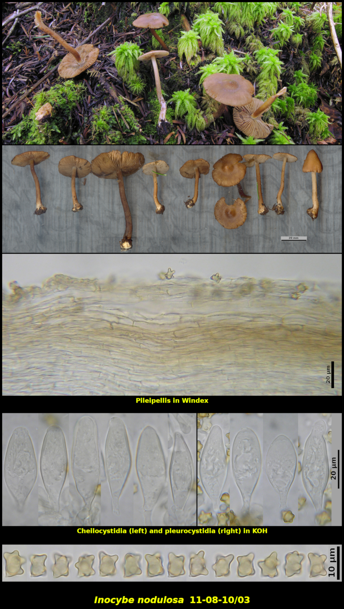
Gregarious in soil with a light Sphagnum cover in wet drainage area under Abies balsamea, Little Lepreau, New Brunswick (11-08-10/03).
Basidiospores reddish brown in spore print, with 8-9 large nodules, 6.4-8.5 x 4.7-7.5 μm, Q = 0.97-1.54 (average[38]: 7.5 x 6.1 μm, Q = 1.25). Cheilocystidia ventricose to subcylindrical, mostly thin-walled, with walls up to 1-2 μm thick in the upper part, hyaline in KOH, with or without crystals at the apex, 41-49 x 12-18 μm. Pleurocystidia abundant, similar to the cheilocystidia, 33-54 x 14-16 μm. Pileipellis a cutis, pale, seated over a brown subpellis of short broad hyphae.
Inocybe nodulosa is very close in appearance to I. napipes, leading to the possiblity that they are in fact one species, with I. napipes the older (1917 vs. 1924) and thus correct name. However, Dr. Brandon Matheney, working in the southern Appalachian Mountains of Tennessee and North Carolina has collected material he believes to represent each of the two species and has deposited DNA sequences to support this. In Version 04 (23 Dec 2017) of his Key to Species of Inocybe from eastern North America he separates the two species based partly on the shape of the bulb at the base of the stipe (napiform or turnip-shaped for I. napipes: flattened and marginate for I. nodulosa), and growth under hemlock (Tsuga canadensis) for I. napipes and association with other conifers, such as Abies spp., for I. nodulosa.
Recognizing I. nodulosa is not difficult. As it is found in eastern Canada, it grows in wet mossy places, especially in Sphagnum patches in association with fir and spruce. The stipe is smooth and lacks cystidia and has a prominant flattened bulb at its base. The cap is dark brown and rather smooth. The flesh lacks a characteristic odour. We have studied material from New Brunswick, Newfoundland and Labrador and Ontario, all of which match these characteristics closely. They are also all similar microscopically, having remarkably nodulose basidiospores and mostly thin-walled cystidia.
Photo: D. Malloch (11-08-10/03).