Fleshy Fungi of New Brunswick >>
Inocybe lacera
Inocybe lacera (Fr.) P. Kumm.
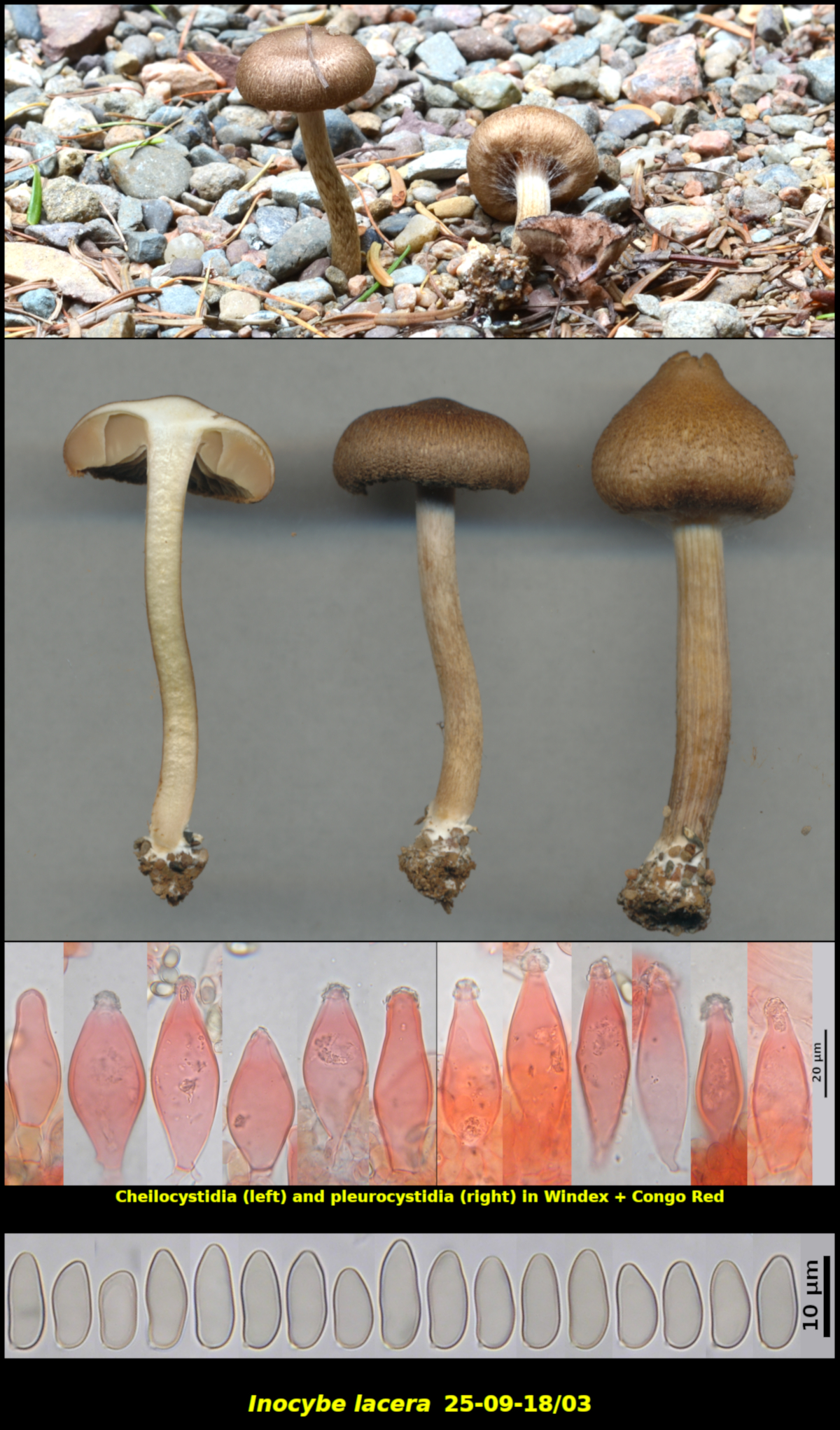
Scattered (2) at the edge of a gravel driveway, associated with Abies balsamea, Picea mariana and Larix laricina – 1.5 km west of Little Lepreau, New Brunswick (25-09-18/03).
Pileus convex-turbinate to convex, with a low to prominent umbo, dry, finely radially fibrillose, not rimose, densely covered with minute subappressed scales, slightly shiny, orange brown (HSV45:70:40-60), 15-17 mm in diameter. Stipe equal, only very slightly enlarged at the base, densely banded with remnants of the universal veil, nearly white at the apex, dull orange brown (HSV40:40:50-70) below, darkening further during maturation, white at the base, dry, 32-40 X 3 mm. Lamellae light brown (HSV35:50:60-70), close, adnexed, not marginate. Cortina conspicuous, white, joining the stipe near its apex. Flesh white in the pileus, dull yellow (HSV45;10:100) in the stipe, with a spermatic odour.
Basidiospores not producing a visible spore print, narrowly ellipsoidal or ovoid to almost boletoid, smooth, without nodules, 9.6-13.5 x 4.6-5.5 μm, Q = 1.94-2.65 (average[39]: 11.4 x 5.0 μm, Q = 2.28). Cheilocystidia forming a continuous sterile margin, fusoid to ventricose, with apex rather tapered and only very rarely swollen, moderately thick-walled, less commonly thin-walled, with walls up to 4.3 µm thick near the apex, usually with apical crystals, 39-57 x 15.1-26.3 μm. Pleurocystidia scattered but abundant, fusoid to ventricose, with apex rather tapered and only very rarely swollen, moderately thick-walled, less commonly thin-walled, with walls up to 2.4 µm thick near the apex, usually with apical crystals, 47-71 x 12.7-22.5 μm.
Inocybe lacera is probably the most common species of Inocybe in New Brunswick. It is usually found in sandy or gravelly disturbed areas along old roads or clearcuts. It seems to form mycorrhizae with a variety of trees, mostly conifers or birch in our area and can be collected in the spring when few other mycorrhizal fungi are in evidence.
Recognizing I. lacera is not difficult, at least if you examine it with a microscope. The remarkably long basidiospors have an aspect ratio (Q-value) generally greater than 2.0. The cystidia are usually rather tapered toward the apex, have variously thickened walls and crystals at the apex. There is a brief discussion and illustration of another New Bruswick Collection of this species (05-06-04/01) in the Essays section of this website.
Photo: D. Malloch (11-08-10/03).