Fleshy Fungi of New Brunswick >>
Hydropus_marginellus
Hydropus_marginellus (Pers.) Singer


Two collections:
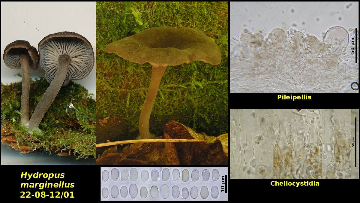
1. Solitary to clustered on a rotting conifer log under Picea rubens, Abies balsamea and Betula papyrifera, Caledonia Gorge Protected Natural Area, New Brunswick (22-08-12/01).
Basidiospores ellipsoidal to slightly phasaeoliform, hyaline, amyloid, 6.2-7.5 X 3.8-4.6 μm, Q = 1.5-1.8 (average[12]: 6.9 x 4.1 μm, Q = 1.67). Cheilocystidia abundant, lanceolate to ventricose-rostrate. Pleurocystidia lacking (or maybe rare). Pileipellis a hymenoderm with brown cells. Pilocystidia abundant. Caulocystidia abundant, fasiculate, similar to the cheilocystida but less regular in shape
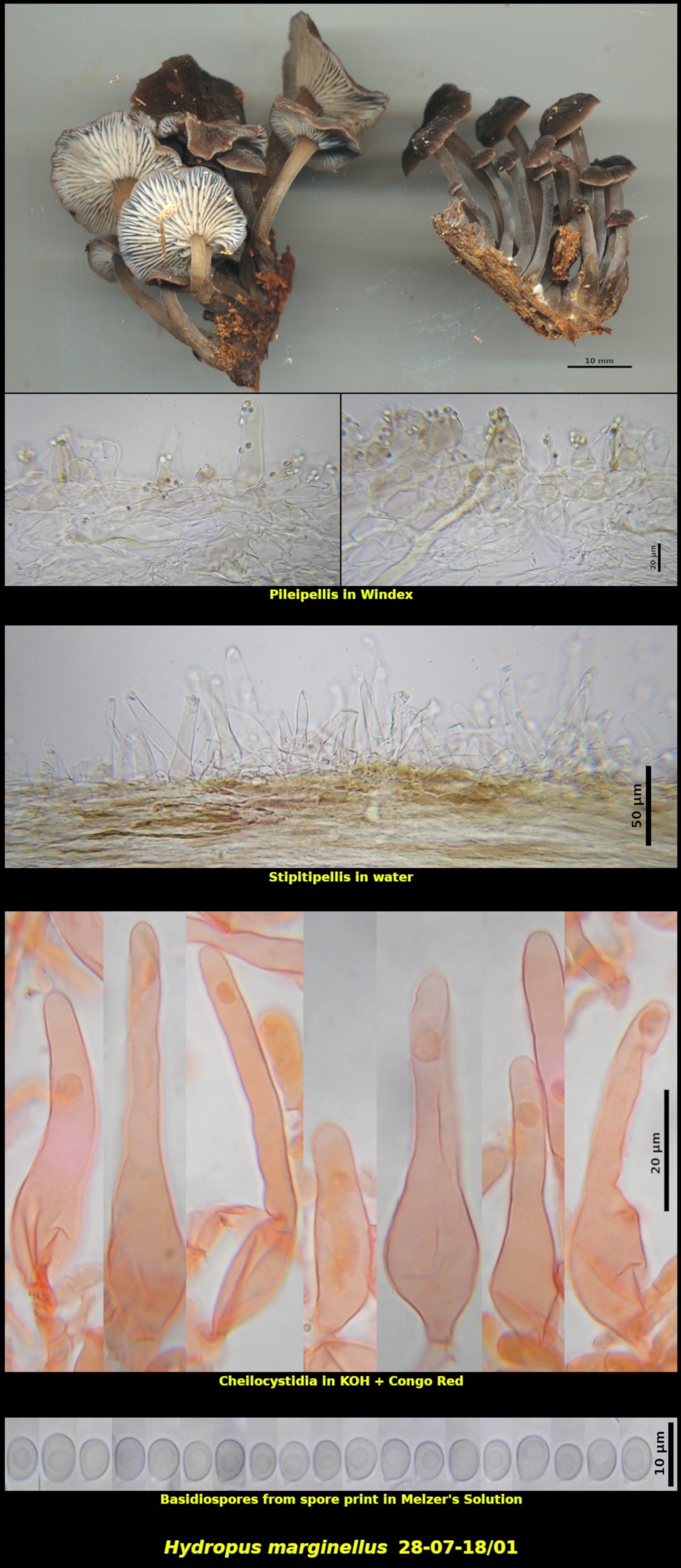
2. Clustered (several) on dead wood in a hollow at the base of a standing Abies balsamea currently occupied by porcupines – Little Lepreau, New Brunswick (28-07-18/01).
Basidiospores white in spore print, ellipsoidal, smooth-walled, strongly amyloid, 5.7-7.5 x 4.2-5.2 μm, Q = 1.25-1.48 (average[33]: 6.4 x 4.7 μm, Q = 1.37). Basidia 4-spored, clamped at the base. Cheilocystidia abundant and forming a sterile margin, ventricose-rostrate, thin-walled, 36-76 x 10.3-19.1 μm. Pleurocystidia not found. Pileipellis a loose hymenoderm of broad clavate to ventricose cells, with many of the cells prolonged into long necks and thus resembling the cheilocystidia, with cells containing a brown intracellular pigment, with subpellis a parallel layer of broad hyphae 2-3 cells deep.
Species of Hydropus are often characterized by the clear liquid that emerges from small cuts in the stipe or pileus. However, the field notes of both of these collections make no mention of this liquid, so, if present, it must not be conspicuous. The abundant lactiferous hyphae in the tissues may be the source of the liquid. Hydropus marginellus is recognized in the field by its brown velvety caps and stipes, pale gills, brown-marginate lamellae and growth on rotting wood of conifers. Microscopically, the amyloid basidiospores and large cheilo-, pilo- and caulocystidia are diagnostic.
The two collections here are nearly identical, differing only in the shape and width of their basidiospores. The brown margins of the lamellae, usually thought to be characteristic of the species, are inconspicuous and could be easily overlooked.
Photo: D. Malloch (22-08-12/01, 28-07-18/01).