Fleshy Fungi of New Brunswick >>
Cortinarius chrysolitus
Cortinarius chrysolitus Kauffman
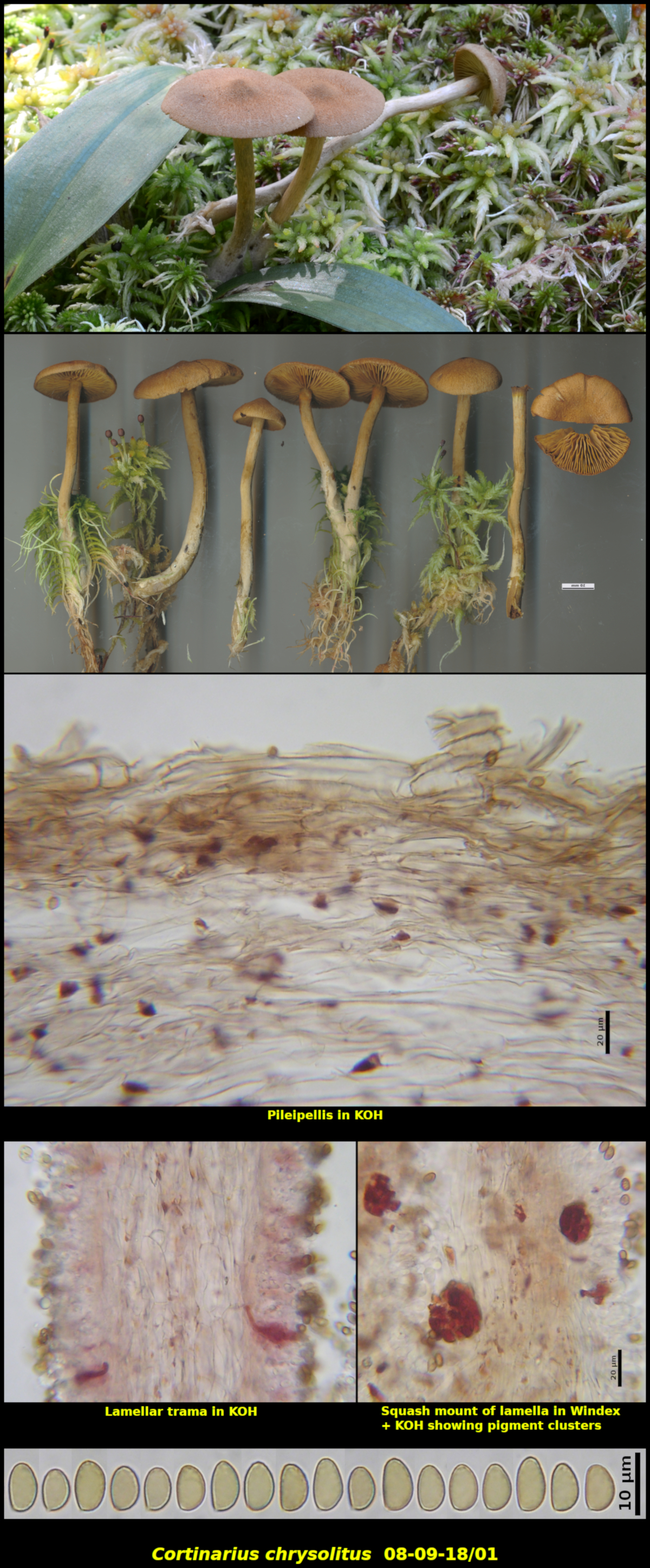
Scattered to gregarious (8) in a deep Sphagnum carpet, in perhumid forest, associated with Abies balsamea and Betula cordifolia, Little Lepreau, New Brunswick (08-09-18/01).
Pileus conic-convex at first, expanding to more broadly so at maturity, with a low but prominent umbo, dry, very finely tomentose-scaly giving the surface a sandpaper-like texture, matte, yellow brown (HSV40;30-40:70-80), 18-36 mm in diameter – dark reddish brown (HSV25:80:30-40) in KOH. Stipe equal, often slightly tortuous, glabrous except for the base which is white-tomentose where in contact with the substrate, dull greyish yellow (HSV55:10-15:80-90), dry, 72-80 X 3-5 mm – with surface instantly dull reddish brown in KOH but rapidly fading; stipe context unchanging in KOH. Lamellae yellow (HSV55;20-30:80-90), close, adnexed, not marginate. Cortina not seen. Flesh nearly concolorous with the lamellae in the area just below the pileipellis, greyish olive (HSV55:40:60) in the rest of the pileus, concolorous with the surface tissues in the stipe, with a nondescript odour and taste – darkening in KOH.
Basidiospores reddish brown in spore print. narrowly obovoid, finely roughened, darkening slightly in Melzer’s Solution, 6.8-8.6 x 4.4-5.1 µm, Q = 1.50-1.84 (average[30]: 7.7 x 4.7 µm, Q = 1.65). Trama of pileus and lamellae turning pale purple in 3% KOH, with numerous dark orange crystals and crystalline masses that turn purplish red in 3% KOH.
Cortinarius chrysolitus is almost always found growing among gametophytes of Sphagnum spp. in coniferous forests. It is most often recognized by the rather bright yellow-green to olive colours or its lamellae and flesh and its long stipe that arises deeply within the moss carpet. It is known from northern regions of North America and Europe.Collection 08-09-18/01 has rather small basidiospores compared to other collections of this species, however, thanks to Dr. J. F. Ammirati, its ITS region has been sequenced and it fits quite well within the species.
Photograph: D. Malloch (08-09-18/01).